
Fig. 2
- ID
- ZDB-IMAGE-110622-87
- Genes
- Publication
- Yang et al., 2011 - BMP and non-canonical Wnt signaling are required for inhibition of secondary tail formation in zebrafish
- All Figures
- Figures for Yang et al., 2011
|
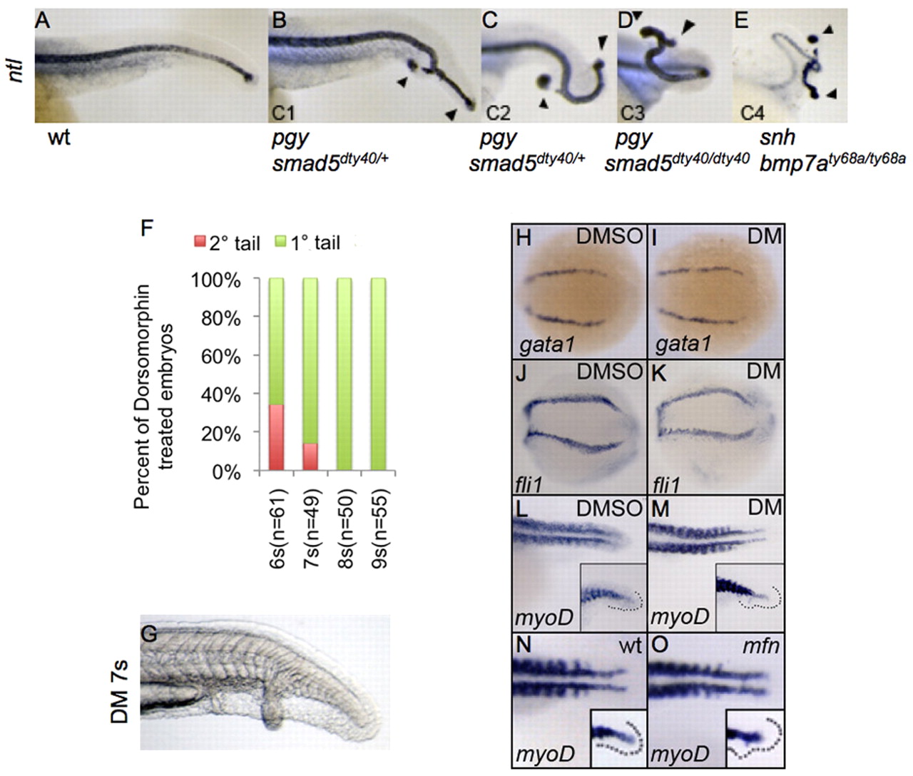
Fig. 2
Secondary tail formation is independent of the role of BMP signaling in fate patterning. (A-E) Lateral view of zebrafish tails in wild-type and dorsalized embryos of indicated genotypes at 24 hpf, after whole-mount in situ for ntl. Primary and secondary tails are marked with arrowheads. C1-C4 refer to the degree of dorsalization using the dorso-anterior index. (F) Distribution of secondary tail phenotype in embryos following dorsomorphin (DM) treatment at the indicated time points. Secondary tails were scored by the presence of ectopic ntl expression. (G) Lateral view of the tail of a live embryo at 24 hpf, after DM treatment at 7 somites (7s). Note the secondary tail with fully developed ventral fin. (H-M) Dorsal view of embryos expressing gata1 (H,I), fli1 (J,K) at the 13-somite stage and myoD (L,M) at 24 hpf in the tail, after treatment with DMSO (H,J,L) and DM (I,K,M) at the 5-somite stage. (N,O) Dorsal view of myoD expression in the tail of wild-type (wt; J) and mfn (K) embryos at 22 hpf. Inset shows the lateral view. Expression of gata1 (I), fli1 (K) and myoD (M,O) is unaffected.