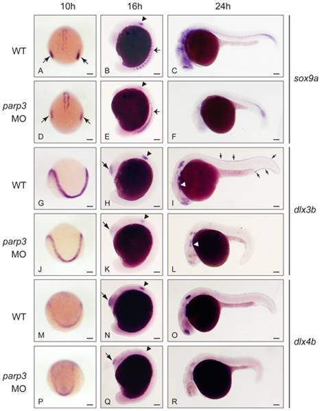
Fig. 6 Impaired expression of sox9a, dlx3b and dlx4b in parp3 morphants.
Zebrafish embryos were untreated (WT) or were injected with 4 ng parp3 MO1. Gene expression was detected by in situ hybridization. A–F. The expression of sox9a is drastically reduced in the otic placodes (small arrows) at 10 hpf and in the otic vesicles (arrowheads) at 16 hpf. Expression of sox9a in somite cells (small arrows in B and E) appears diffuse in parp3 morphants. Expression of sox9a is almost completely abolished in the head region at 24 hpf (C, F). Expressions of dlx3b (G–L) and dlx4b (M–R) are minimally affected by parp3 MO in ectodermal cells at 10 hpf (G, J, M, P) but are significantly reduced in the otic vesicles (arrowheads), olfactory placodes (large arrows) and branchial arches (white arrows) of parp3 morphants at 16 hpf (H, K, N, Q) and 24 hpf (I, L, O, R). The expression of dlx3b and dlx4b is abolished in the median fin fold of 24 hpf parp3 morphant embryos (small arrows in I). Dorsal views of embryos with anterior to the bottom in A, D, G, J, M, P and lateral views with anterior to the left, dorsal to the top, in B, C, E, F, H, I, K, L, N, O, Q and R. Scale bars represent 10 μm.

