Image
Figure Caption
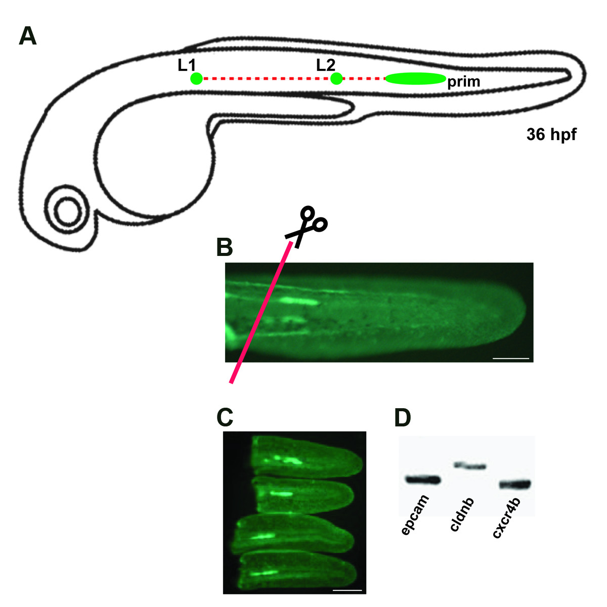
Fig. 1 Section of the transgenic line cldnb:gfp. (A) Schematic drawings of a migrating primordium of the posterior lateral line along the horizontal myoseptum at 36 hpf. (B) Transgenic cldnb:gfp embryo at 36 hpf, when tails were sectioned (red line). (C) Sectioned tails with GFP primordium. (d) RT-PCR of RNA derived from dissected tails of 36 hpf embryos with primer specific to three genes known to be expressed in the primordium at this stage. L1: neuromast 1, L2: neuromast 2, prim: LLP primordium. Scale bars are 50 μm in (b-c).
Figure Data
Acknowledgments
This image is the copyrighted work of the attributed author or publisher, and
ZFIN has permission only to display this image to its users.
Additional permissions should be obtained from the applicable author or publisher of the image.
Full text @ BMC Dev. Biol.

