
Fig. 3
- ID
- ZDB-IMAGE-101104-53
- Genes
- Publication
- Aamar et al., 2010 - Sox17 and chordin are required for formation of Kupffer's vesicle and left-right asymmetry determination in zebrafish
- All Figures
- Figures for Aamar et al., 2010
|
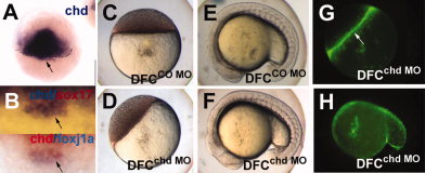
Fig. 3 Chordin expression in the dorsal forerunner cells in zebrafish embryos and targeting of DFC by chd MO. A,B: Whole mount in situ hybridization for chd at 50% epiboly (A), and double staining of chd (blue) and sox17 7 (red) (B, top), and chd (red) and foxj1a (blue) (B, bottom) at 60% epiboly; arrows point to DFC. Embryos were injected at the midblastula stage with 2.5 ng CO MO (C,E) or chd MO (D,F), and photographed at the oblong (C,D) and 16-18 somite (E,F) stages. The embryos in D,F are also shown as fluorescent images (G,H, respectively). Arrow in G, yolk syncycial layer. A,B, dorsal views; E,F,H, lateral views, with anterior to the left.