
IMAGE
Fig. 4
- ID
- ZDB-IMAGE-101012-17
- Genes
- Publication
- Mei et al., 2010 - Mtmr8 is essential for vasculature development in zebrafish embryos
- All Figures
- Figures for Mei et al., 2010
Image
|
Figure Caption
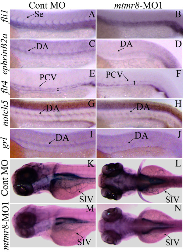
Fig. 4 The effects of mtmr8 knockdown on vascular marker genes and vascular endothelium. (A-J) Whole mount in situ hybridization with the vascular marker gene-specific probes (as shown on the left) in Cont-MO (A, C, E, G and I) and mtmr8-MO (B, D, F, H and J) embryos at 26 hpf. The signals of DA, PCV and Se are indicated by arrows, and the PCV expansion size is indicated by double-arrowheads in the flt4 probe hybridization (E and F). (K-N) Lateral (K, M) and dorsal (L, N) views of alkaline phosphatase-stained Cont-MO (K, L) and mtmr8-MO (M, N) embryos at 3 dpf.
Figure Data
Acknowledgments
This image is the copyrighted work of the attributed author or publisher, and
ZFIN has permission only to display this image to its users.
Additional permissions should be obtained from the applicable author or publisher of the image.
Full text @ BMC Dev. Biol.