
Fig. 2
- ID
- ZDB-IMAGE-100816-13
- Genes
- Publication
- Goody et al., 2010 - Nrk2b-mediated NAD+ production regulates cell adhesion and is required for muscle morphogenesis in vivo: Nrk2b and NAD+ in muscle morphogenesis
- All Figures
- Figures for Goody et al., 2010
|
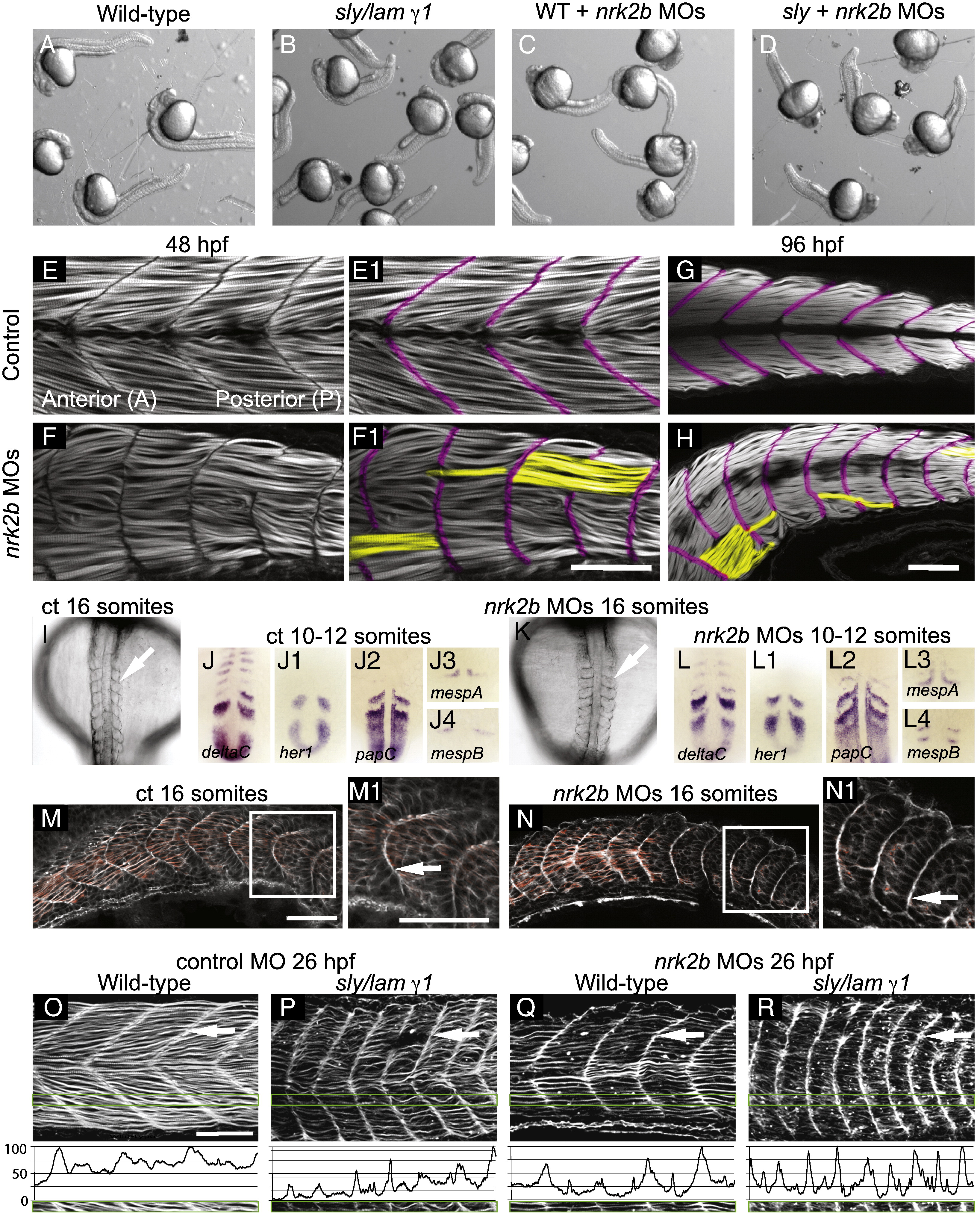
Fig. 2 Disrupted MTJs in nrk2b morphants are not due to discontinuous initial somite boundaries. (A–D) Brightfield images of a dish of 26 hpf embryos. (A) Wild-types. (B) sly/laminin γ1 mutants. (C) nrk2b morphants. (D) nrk2b MO-injected-laminin γ1 mutants. Note that injection of nrk2b MOs into laminin mutants does not drastically worsen their phenotype or result in death. (E–H) Side mount, anterior left, dorsal top, phalloidin stained embryos, MTJ boundaries are pseudocolored fuchsia and abnormally long muscle fibers are pseudocolored yellow. (E–E1) 48 hpf control. (F–F1) 48 hpf nrk2b morphant (71/212, 33% of MTJs are crossed by muscle fibers). (G) 96 hpf control. (H) 96 hpf nrk2b morphant (19/67, 28% of MTJs are crossed by muscle fibers). (I, K) Dorsal mount, anterior top, 16 S, brightfield images. Note that somite boundaries form (white arrows) in nrk2b morphants (K). (Panels J and L) Dorsal mount, anterior top, 10–12 S, ISH for known somite patterning and somite boundary formation genes. Note that the domains and patterns of expression are similar between controls (panel J) and nrk2b morphants (panel L). (M–N) Side mount, anterior left, dorsal top, 16 S, phalloidin staining. Note concentrated actin at the initial somite boundary (white arrows). These data show that discontinuous MTJs are not due to discontinuous initial somite boundaries. (O–R) Side view, anterior left, dorsal top, 26 hpf embryos, phalloidin stained to visualize actin. (O–P) Control MO-injected. (Q–R) nrk2b MO-injected. (O, Q) Wild-types. (P, R) sly/laminin γ1 mutants. Note that in laminin mutants and nrk2b morphants, actin is enriched at the MTJ (white arrows) and lacking in the myotome. Green boxes correspond to normalized fluorescence intensity (see Materials and methods) plots. These graphs support the qualitative actin disruption seen in laminin mutants and nrk2b morphants. Scale bars are 50 μm.
Reprinted from Developmental Biology, 344(2), Goody, M.F., Kelly, M.W., Lessard, K.N., Khalil, A., and Henry, C.A., Nrk2b-mediated NAD+ production regulates cell adhesion and is required for muscle morphogenesis in vivo: Nrk2b and NAD+ in muscle morphogenesis, 809-826, Copyright (2010) with permission from Elsevier. Full text @ Dev. Biol.