
IMAGE
Fig. S2
- ID
- ZDB-IMAGE-100225-7
- Genes
- Publication
- Sun et al., 2010 - Zili Inhibits TGF-beta Signaling by Interacting with Smad4
- All Figures
- Figures for Sun et al., 2010
Image
|
Figure Caption
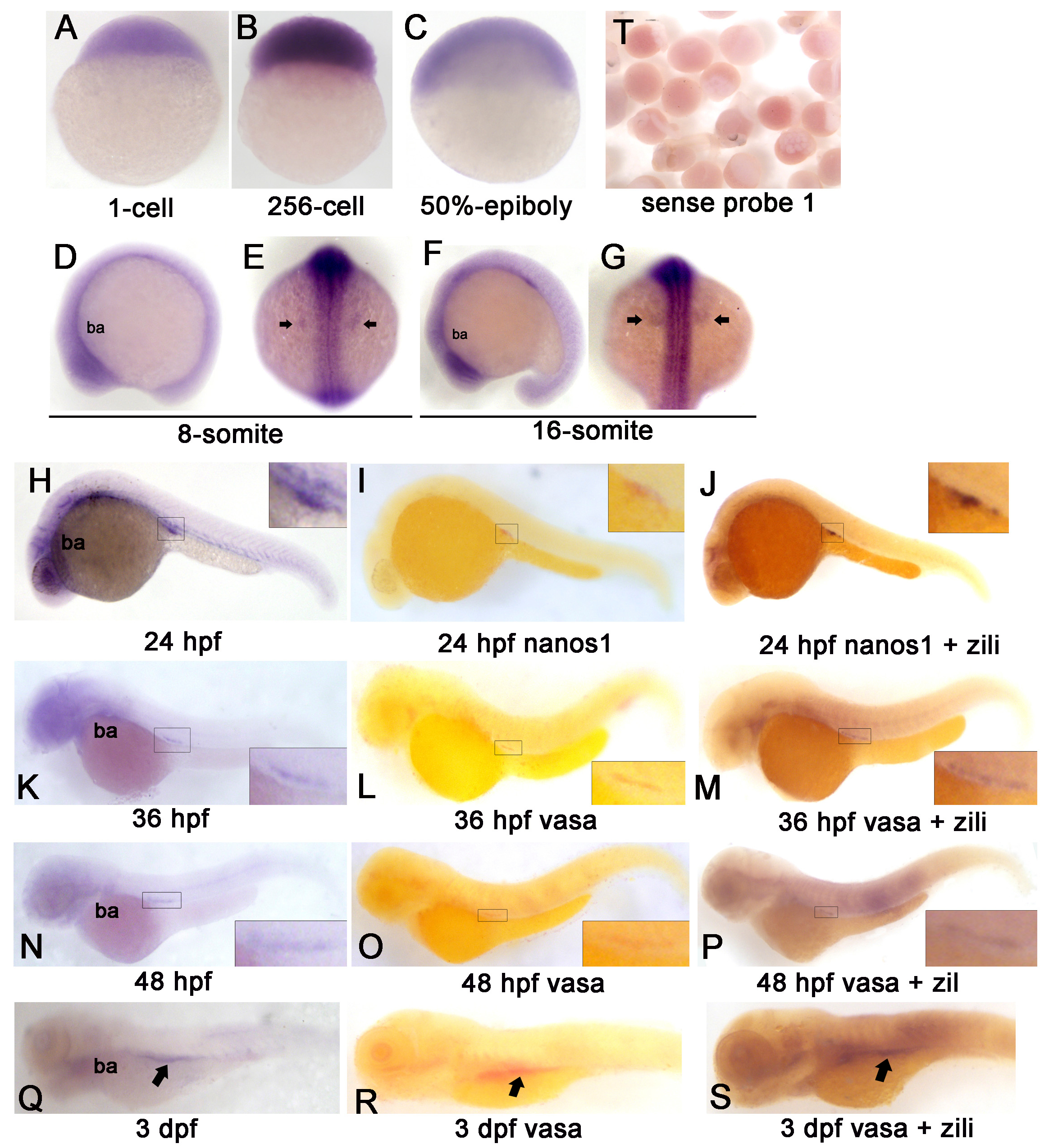
Fig. S2 Spatiotemporal Expression Pattern of zili transcript in zebrafish embryos at indicated stages. Detection by whole-mount in situ hybridization (Probe 1). Embryo orientations: (A–C) lateral views with the animal pole oriented at the top; (E and G) dorsal views with anterior oriented at the top; (D, F, H-S) lateral views with anterior oriented toward the left. The indicated domains: ba, branchial and pharyngeal arches. PGCs region is enlarged in the insert in (H-S) and indicated by arrow in (E, G, Q-S). (T) Embryos detected by sense probe 1.
Figure Data
Acknowledgments
This image is the copyrighted work of the attributed author or publisher, and
ZFIN has permission only to display this image to its users.
Additional permissions should be obtained from the applicable author or publisher of the image.
Open Access.
Full text @ J. Biol. Chem.