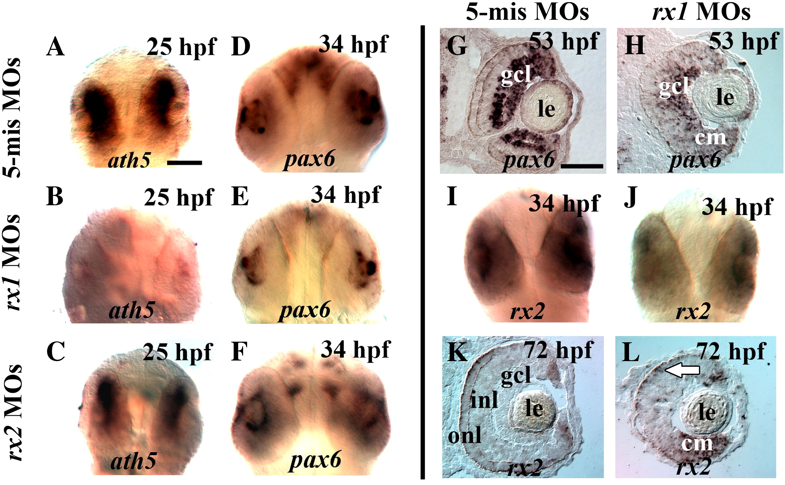
Fig. 4 Expression of ath5 and pax6 but not rx2 is disrupted in rx1-depleted embryos. (A–C) Embryos fixed at 25 hpf and processed for in situ hybridization with an ath5 probe show strong retinal labeling in control embryos (A), no labeling in rx1-depleted embryos, and a normal ath5 expression pattern in rx2-depleted embryos. (D–F) Embryos fixed at 34 hpf and processed for in situ hybridization with a pax6 probe, showing labeling in the retina, lens and forebrain of control embryos (D), labeling in lens and forebrain only in rx1-depleted embryos (E), and a normal pattern of expression of pax6 in rx2-depleted embryos (F). (G–H) Sections obtained from embryos fixed at 53 hpf following injection with control-MOs (G) or rx1-depleting MOs (H) and hybridized with a probe for pax6, showing pax6 expression in the retinas of the morphants, albeit in a pattern reflecting a delay in neurogenesis. (I–L) Embryos processed at 34 hpf as whole mounts (I, J) or at 72 hpf as cryosections (K, L), for in situ hybridization with an rx2 probe, following injection with control-MOs (G) or rx1-depleting MOs, showing that rx2 is not regulated by rx1. le = lens; gcl = ganglion cell layer; inl = inner nuclear layer; onl = outer nuclear layer; cm = ciliary margin; scale bar in A and G = 50 μm.
Reprinted from Developmental Biology, 328(1), Nelson, S.M., Park, L., and Stenkamp, D.L., Retinal homeobox 1 is required for retinal neurogenesis and photoreceptor differentiation in embryonic zebrafish, 24-39, Copyright (2009) with permission from Elsevier. Full text @ Dev. Biol.

