
Fig. 6
|
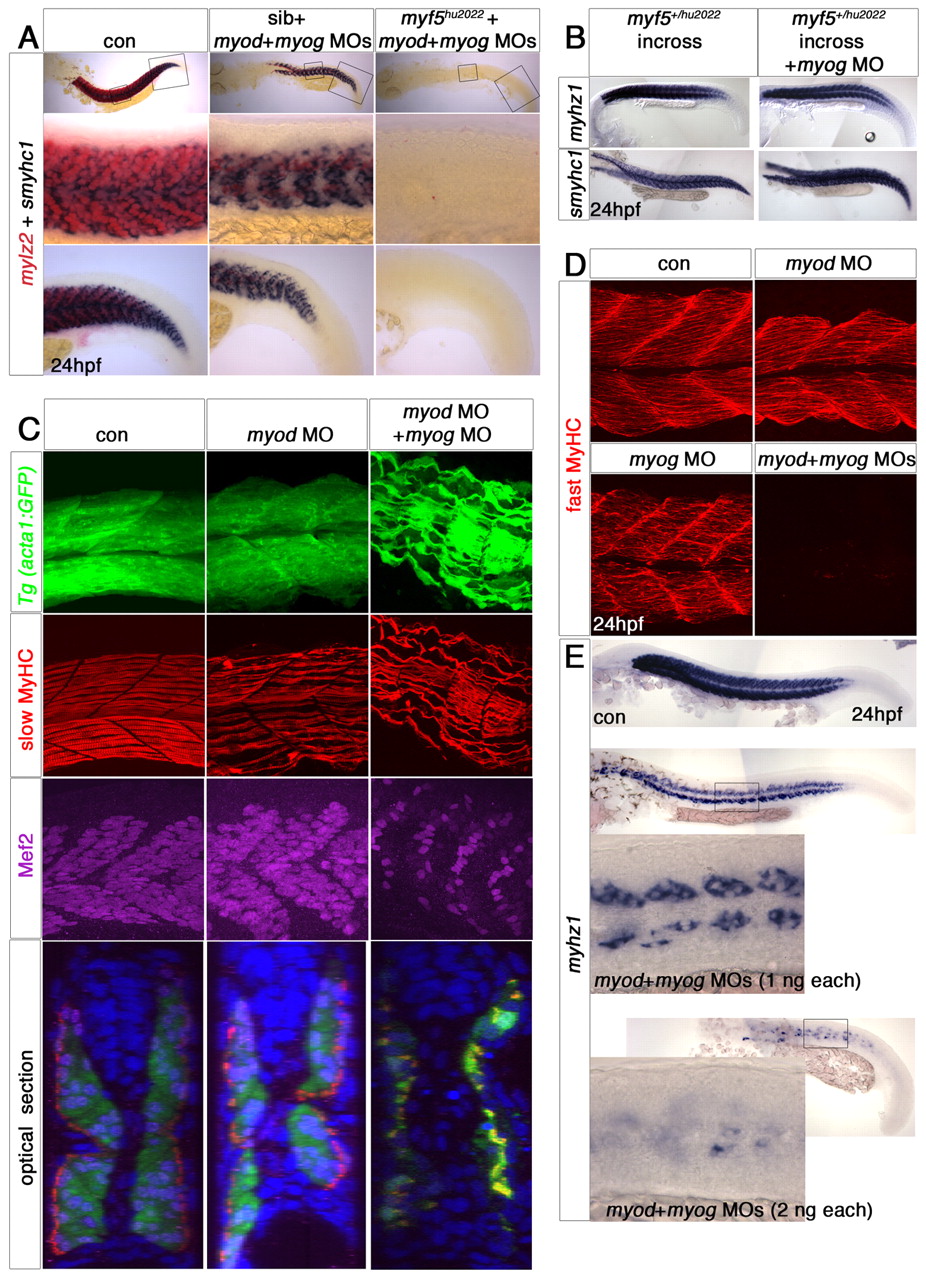
Fig. 6 Loss of Myod and Myog ablates somitic fast muscle. In situ mRNA hybridisation (A,B,E) or immunofluorescent confocal stacks (C,D) of 24 hpf wild-type (con), Tg(acta1:GFP) or myf5+/hu2022 incrosses injected with the indicated MOs. Lateral views, anterior to left. (A) Magnified regions (boxed) showing that lack of Myf5, Myod and Myog results in loss of all muscle. Knockdown of Myod and Myog results in loss of most fast, but not slow, muscle. Note that mylz2 (red) is weakly expressed in immature slow muscle. (B) Loss of Myog and Myf5 has little effect, if any, on fast myhz1 or slow smyhc1 expression. (C) Injection of myod+myog MOs into Tg(acta1:GFP) fish results in loss of fast muscle, as shown by loss of Mef2 staining and GFP medial to the slow fibre layer accompanied by somitic apoptosis as revealed with DAPI (blue in the optical transverse sections). Elongated mononucleate slow fibres differentiate and express GFP, slow MyHC and Mef2. myod morphants confirm the lateral reduction of fast muscle mass. (D) myod+myog MOs ablate fast MyHC in somites, whereas single MOs have a lesser effect. (E) Titration of myod+myog MOs progressively diminishes residual myhz1-expressing cells. The lower panels are high-magnification images of mid-trunk somites (boxed).