
Fig. 8
- ID
- ZDB-IMAGE-090127-15
- Genes
- Antibodies
- Publication
- Noël et al., 2008 - Organ-specific requirements for Hdac1 in liver and pancreas formation
- All Figures
- Figures for Noël et al., 2008
|
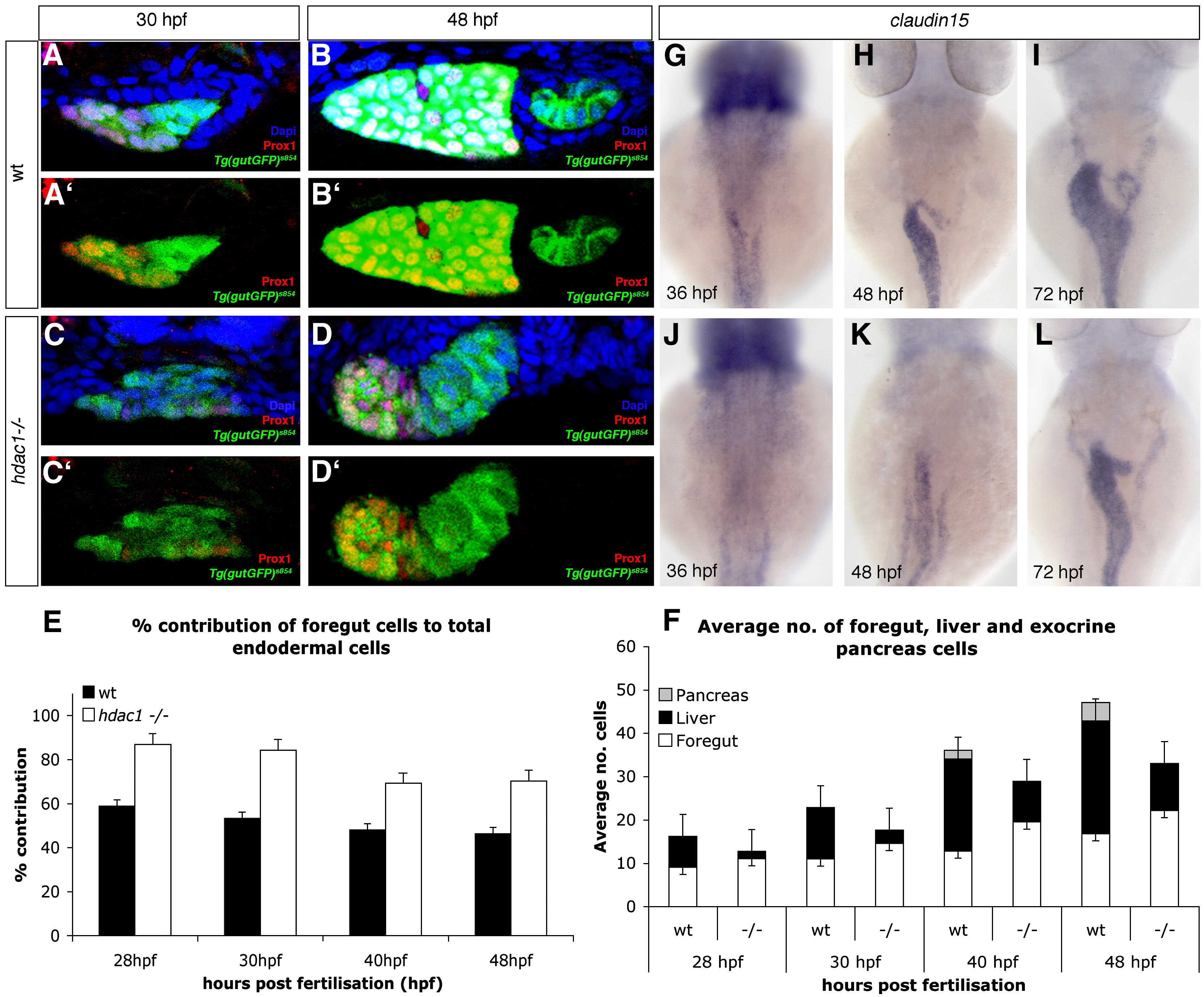
Fig. 8 Loss of Hdac1 function results in an increase of non-hepatic foregut endoderm. (A–D) Transverse sections taken through the level of the liver, at 30 hpf and 48 hpf in wt (A–B′) and hdac1 mutants (C–D′). To count endodermal cells, Tg(gutGFP)s854 embryos were stained for Prox1 (red), highlighting hepatoblasts, and Dapi (blue). (E) hdac1 mutants display an increased percentage contribution of foregut cells to total endodermal cells between 28 hpf and 48 hpf when compared to wild type siblings. (F) hdac1 mutant embryos have an increased number of foregut cells compared to wild type embryos between 28 and 48 hpf (white bars). However, hdac1 mutant embryos display an overall reduction in number of total endodermal cells when compared to wild type embryos (compare total bar height). (E, F) Number of wild type embryos analysed: 28 hpf n = 12, 30 hpf n = 14, 40 hpf n = 13, 48 hpf n = 13. Number of hdac1 mutants analysed: 28 hpf n = 10, 30 hpf n = 11, 40 hpf n = 10, 48 hpf n = 10. (G–L) hdac1 is required for correct temporal expression of claudin15. Wild type embryos express claudin15 in the endoderm from 36 hpf onwards (G–I), whereas hdac1 mutants fail to express claudin15 at 36 hpf (J), however, expression is detected at 48 hpf (K) and 72 hpf.
Reprinted from Developmental Biology, 322(2), Noël, E.S., Casal-Sueiro, A., Busch-Nentwich, E., Verkade, H., Dong, P.D., Stemple, D.L., and Ober, E.A., Organ-specific requirements for Hdac1 in liver and pancreas formation, 237-250, Copyright (2008) with permission from Elsevier. Full text @ Dev. Biol.