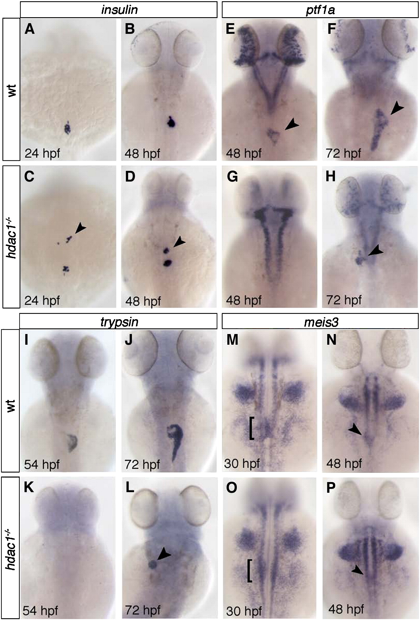
Fig. 7 Hdac1 depletion results in ectopic endocrine tissue formation and defects in exocrine pancreas specification. (A–D) insulin is expressed in a single cluster of endocrine β-cells in wild type embryos at 24 and 48 hpf (A, B). hdac1 mutants display ectopic anterior clusters of insulin-expressing cells at 24 and 48 hpf (arrowheads, C, D). (E–H) ptf1a is expressed in the exocrine pancreas at 48 and 72 hpf (arrowhead, E, F). hdac1 mutant embryos lack pancreatic ptf1a expression at 48 hpf (G), but express ptf1a at 72 hpf in a reduced domain (arrowhead, H). (I–L) trypsin expression in the exocrine pancreas in wild type siblings at 54 hpf (I) is absent in hdac1 mutants (K), however is detected in a subset of hdac1 mutants at 72 hpf (arrowhead, L). (M–P) Wild type embryos express meis3 medially in the LPM at 30 hpf (bracket, M) and 48 hpf (arrowhead, N). hdac1 mutants display meis3 expression bilateral to the endoderm at 30 hpf (bracket, O) and medially at 48 hpf (P).
Reprinted from Developmental Biology, 322(2), Noël, E.S., Casal-Sueiro, A., Busch-Nentwich, E., Verkade, H., Dong, P.D., Stemple, D.L., and Ober, E.A., Organ-specific requirements for Hdac1 in liver and pancreas formation, 237-250, Copyright (2008) with permission from Elsevier. Full text @ Dev. Biol.

