
Fig. 1
- ID
- ZDB-IMAGE-090109-12
- Genes
- Publication
- Linville et al., 2009 - Combinatorial roles for zebrafish retinoic acid receptors in the hindbrain, limbs and pharyngeal arches
- All Figures
- Figures for Linville et al., 2009
|
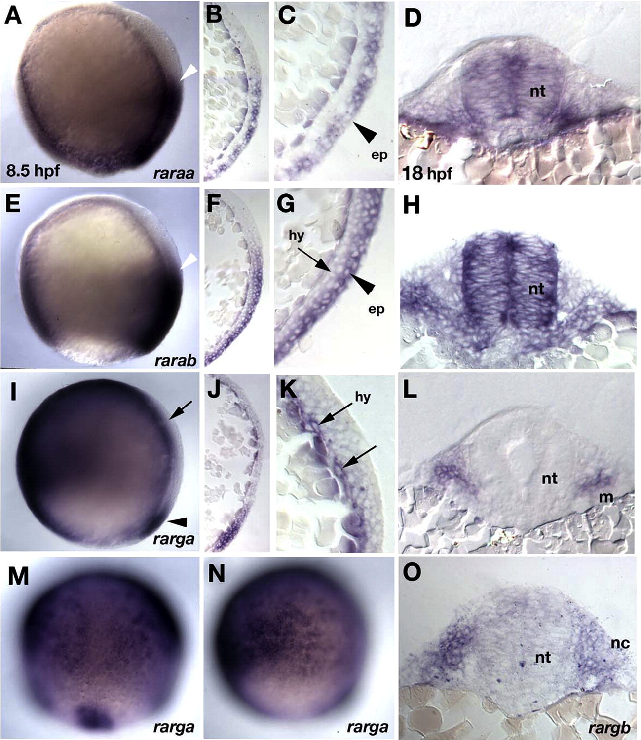
Fig. 1 Germ layer-specific expression of zebrafish rars. In situ hybridization in whole mounts (A, E, I, M, N) and parasagittal sections (B, C, F, G, J, K) at 8.5 hpf (90% epiboly), and transverse sections at 18 hpf (18 somites) at hindbrain levels (D, H, L, O). (A–D) raraa. (A) Expression is in posterior ectoderm in the gastrula, with an anterior border in the presumptive hindbrain (white arrowhead). (B–D) Sections reveal that expression is restricted to epiblast, shown at higher magnification in panel C (arrowhead), and persists in the neural tube (D). (E–H) rarab. (E) Expression is in posterior ectoderm in the gastrula, with an anterior border in the presumptive hindbrain (white arrowhead), similar to raraa. (F–H) Sections reveal strong expression in epiblast (arrowhead in panel G), weaker in hypoblast, which persists in the neural tube and pharyngeal arches (H). (I–N) rarga. (I, M, N) Expressed anteriorly and ventrally during gastrulation and at the dorsal margin (arrowheads in panels I and M). Panel M shows a dorsal view and panel N is focused laterally to show expression in ventral epiblast (presumptive epidermis). (J–L) Sections near the dorsal side reveal expression restricted to the hypoblast (arrows in panel K) and later in cranial mesoderm (L). (O) rargb. Expression is ubiquitous at early stages (not shown), and by 18 hpf sections reveal expression in pharyngeal arch mesenchyme. Abbreviations: ep, epiblast; hy, hypoblast; m, mesoderm; nc, neural crest; nt, neural tube.
Reprinted from Developmental Biology, 325(1), Linville, A., Radtke, K., Waxman, J.S., Yelon, D., and Schilling, T.F., Combinatorial roles for zebrafish retinoic acid receptors in the hindbrain, limbs and pharyngeal arches, 60-70, Copyright (2009) with permission from Elsevier. Full text @ Dev. Biol.