
IMAGE
Fig. S2
- ID
- ZDB-IMAGE-081104-9
- Genes
- Antibodies
- Publication
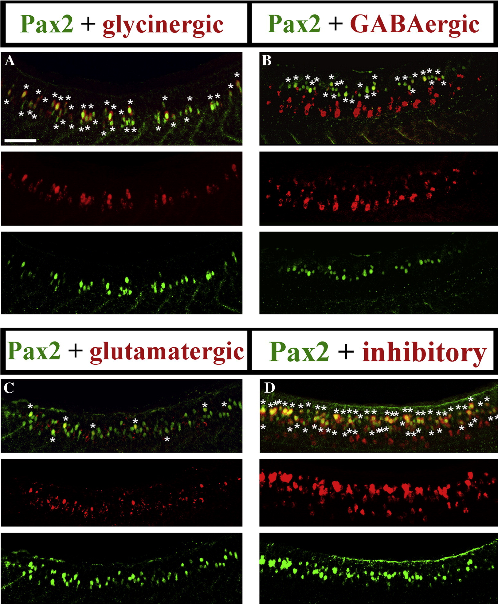
- Batista et al., 2008 - Pax2/8 act redundantly to specify glycinergic and GABAergic fates of multiple spinal interneurons
- All Figures
- Figures for Batista et al., 2008
Image
|
Figure Caption
Fig. S2 Lateral views of Pax2 immunohistochemistry and in situ hybridisation for markers of glycinergic (A), GABAergic (B) glutamatergic (C) and GABAergic or glycinergic (labelled “inhibitory”; probes were mixed) (D) cell fates in 24 h WT trunks. See Materials and methods for details of probes used. The pictures in this figure are the same as in Fig. 2 in the main paper — but this supplementary data figure also shows single channel images for each result. Stars in merged images indicate double labelled cells. Rostal is left, dorsal is top. Scale bar = 50 μm.
Figure Data
Acknowledgments
This image is the copyrighted work of the attributed author or publisher, and
ZFIN has permission only to display this image to its users.
Additional permissions should be obtained from the applicable author or publisher of the image.
Reprinted from Developmental Biology, 323(1), Batista, M.F., and Lewis, K.E., Pax2/8 act redundantly to specify glycinergic and GABAergic fates of multiple spinal interneurons, 88-97, Copyright (2008) with permission from Elsevier. Full text @ Dev. Biol.