
Fig. 2
- ID
- ZDB-IMAGE-081104-5
- Genes
- Antibodies
- Publication
- Batista et al., 2008 - Pax2/8 act redundantly to specify glycinergic and GABAergic fates of multiple spinal interneurons
- All Figures
- Figures for Batista et al., 2008
|
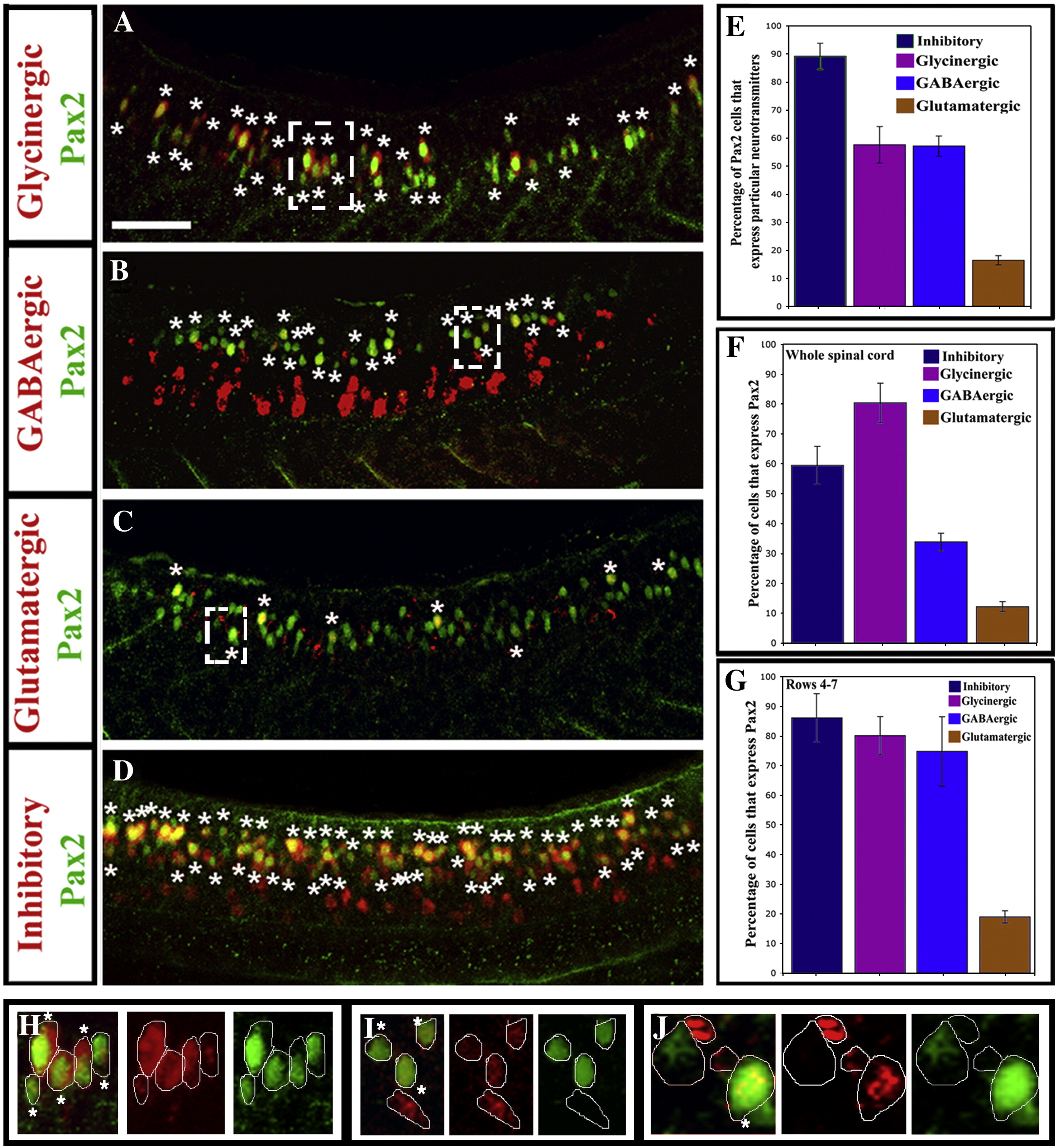
Fig. 2 Most Pax2-expressing zebrafish spinal interneurons are inhibitory. Lateral views of Pax2 immunohistochemistry and in situ hybridisation for markers of glycinergic (A), GABAergic (B) glutamatergic (C) and GABAergic or glycinergic (labelled “inhibitory”; probes were mixed) (D) cell fates in 24 h WT trunks. See Materials and methods for details of probes used. Stars indicate double labelled cells. Rostal is left, dorsal is top. Scale bar = 50 μm. (E) Percentage of Pax2-expressing cells with particular neurotransmitter phenotypes. (F and G) Percentage of cells with specific neurotransmitter phenotypes that express Pax2 in whole spinal cord (F) or just the Pax2-expression domain (rows 4–7; G). In all cases, percentages are averages from 5 different embryos at 24 h. Error bars denote standard deviation. Note that most glycinergic cells are found in the Pax2/8 expression domain but many GABAergic cells are found more ventrally in the spinal cord (A and B; also cf. F and G). (H–J) Magnified views of dashed white boxes in A (H), B (I) and C (J) respectively showing red and green channels and merged images for a single confocal focal plane. Stars in merged images indicate double labelled cells. For single channel images of the whole lateral view see Supp. Data Fig. 2.
Reprinted from Developmental Biology, 323(1), Batista, M.F., and Lewis, K.E., Pax2/8 act redundantly to specify glycinergic and GABAergic fates of multiple spinal interneurons, 88-97, Copyright (2008) with permission from Elsevier. Full text @ Dev. Biol.