
Fig. 4
- ID
- ZDB-IMAGE-081028-14
- Genes
- Antibodies
- Publication
- Ahn et al., 2008 - Tri-phasic expression of posterior Hox genes during development of pectoral fins in zebrafish: Implications for the evolution of vertebrate paired appendages
- All Figures
- Figures for Ahn et al., 2008
|
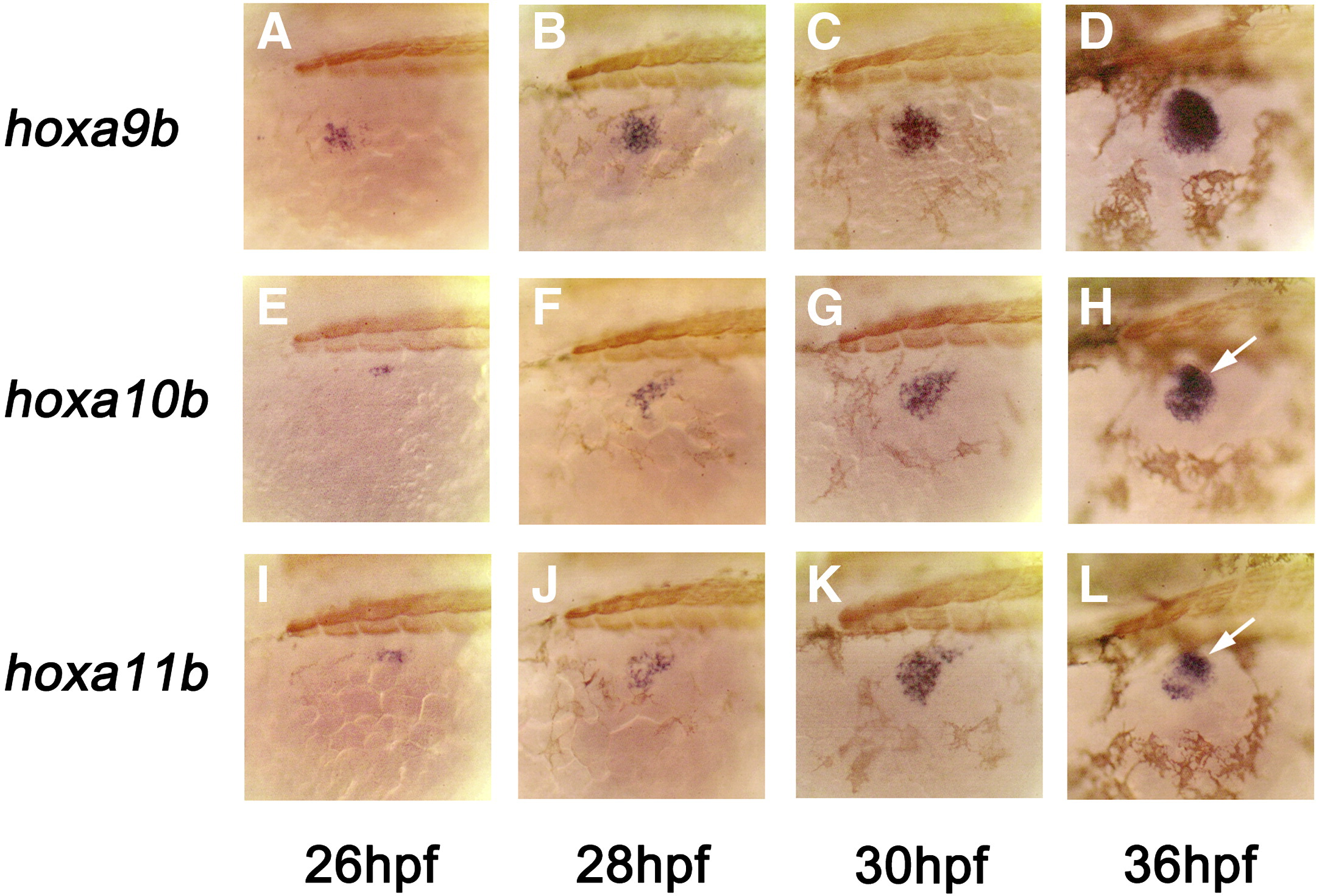
Fig. 4 Early expression of hoxab cluster genes in zebrafish pectoral fins. Expression of hoxa9b starts within the mesenchyme cells of anterior fin field (lying lateral to the second myotome) and subsequently spreads toward the posterior, eventually encompassing the entire fin bud mesenchyme by 30 hpf (A–C: phase I expression). For hoxa10b and hoxa11b, the initial expression is confined to the myogenic mesenchyme cells (E–G, I–K). For these genes, expression within the chondrogenic mesenchyme cells begins around 30 hpf in distal posterior cells (phase II expression) roughly overlying the medial group of myogenic cells (H, L: arrows). In dorsal views, this creates a misleading impression of enhanced expression in the medial cluster of prospective pectoral fin muscle cells (compare Figs. 4H, L with Figs. 2D, I). Oblique dorsal views with anterior to the left in all panels. Each embryo is also counter-stained for muscle myosin (brown) to show the position of myotomes. hpf: hours post fertilization.
Reprinted from Developmental Biology, 322(1), Ahn, D., and Ho, R.K., Tri-phasic expression of posterior Hox genes during development of pectoral fins in zebrafish: Implications for the evolution of vertebrate paired appendages, 220-233, Copyright (2008) with permission from Elsevier. Full text @ Dev. Biol.