Fig. 7
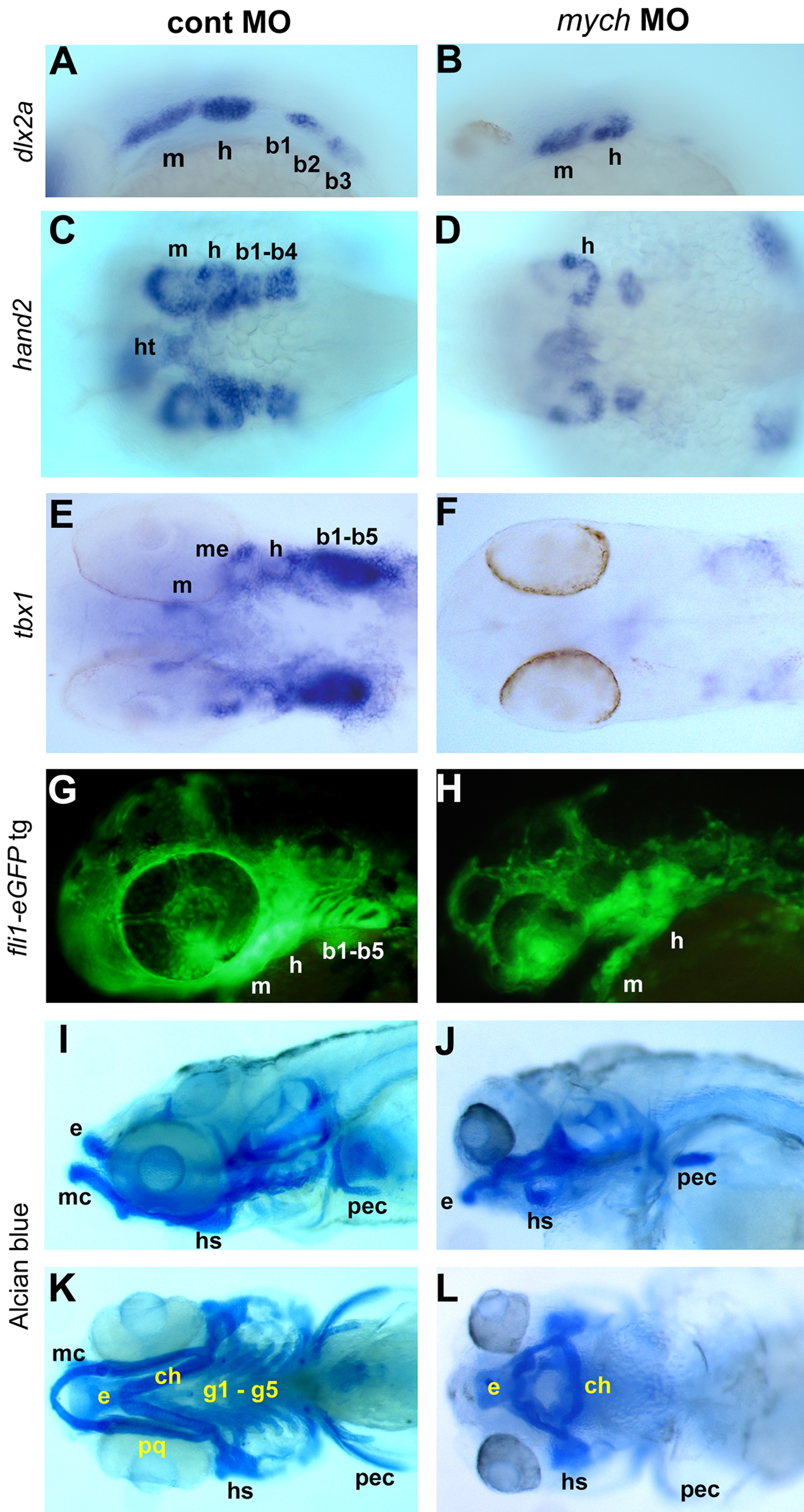
Multiple defects in pharyngeal arch development caused by mych MO.
Control MO (A,C,E,G,I,K) and mych UTR MO-injected embryos (B,D,F,H,J,L). A–B. Lateral view of dlx2a expression in mych UTR MO-injected embryos at 26hpf (B). C–D. Ventral view of expression of hand2 at 36hpf. E–F. Ventral view of tbx1 expression at 45hpf. G–H. Lateral view of fli1-eGFP transgenic line at 40hpf. I–L. Lateral (I,J) and ventral (K,L) views of Alcian blue stained day 5 control (I,K) and mych UTR MO-injected embryos (J,L). ch, ceratohyal, b, branchial arch; e, ethmoid plate; g, gill arches; h, hyoid arch; hs, hyosymplectic; ht, heart; m, mandibular arch; mc, Meckel's cartilage; me, mesoderm; pec, pectoral fin; pq, palatoquadrate.
Image
Figure Caption
Figure Data
Acknowledgments
This image is the copyrighted work of the attributed author or publisher, and
ZFIN has permission only to display this image to its users.
Additional permissions should be obtained from the applicable author or publisher of the image.
Open Access
Full text @ PLoS One

