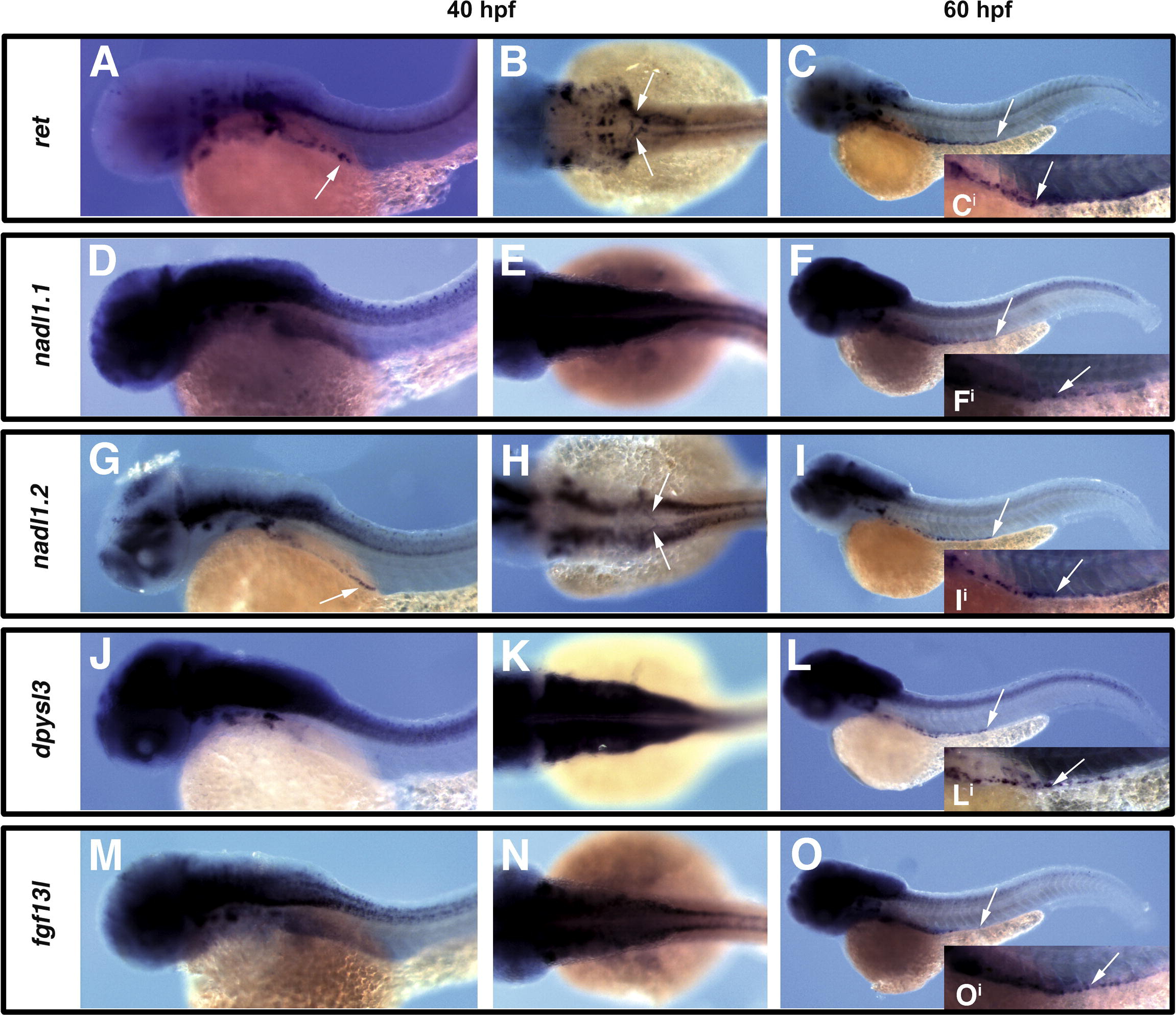
Fig. 5
Fig. 5 Orthologous zebrafish genes are expressed within the ENS and display similar profiles of expression to their mouse counterparts. Analysis of expression of ret (A–C), nadl1.1 (D–F), nadl1.2 (G–I), dpysl3 (J–L), and fgf13l (M–O), within enteric precursors and their derivatives at 40hpf lateral views (A, D, G, J and M) or dorsal views (B, E, H, K and N) and 60hpf lateral views (C, F, I, L and O). Like ret, nadl1.2 is expressed in migratory enteric precursors at earliest stages, as neural crest migrate in two streams towards the centrally located developing gut tube (arrows in A, B, G, H), and persists as enteric neurons are colonizing the gut tube (distal extent of expression within gut tube marked by arrow in C, I, and individual enteric neurons shown in higher magnification in insets Ci and Ii). In contrast, dpysl3, fgf13l, and nadl1.1 are not expressed in early migratory enteric precursors, but are expressed within enteric neurons colonizing the gut tube (see arrows marking distal extent of expression within gut tube in F, L, O, and marking individual enteric neurons in insets Fi, Li, and Oi). Images for comparison are photographed at the same magnification.
Reprinted from Mechanisms of Development, 125(8), Heanue, T.A., and Pachnis, V., Ret isoform function and marker gene expression in the enteric nervous system is conserved across diverse vertebrate species, 687-699, Copyright (2008) with permission from Elsevier. Full text @ Mech. Dev.

