
Fig. 1
- ID
- ZDB-IMAGE-080604-46
- Genes
- Antibodies
- Publication
- Elworthy et al., 2008 - Expression of multiple slow myosin heavy chain genes reveals a diversity of zebrafish slow twitch muscle fibres with differing requirements for Hedgehog and Prdm1 activity
- All Figures
- Figures for Elworthy et al., 2008
|
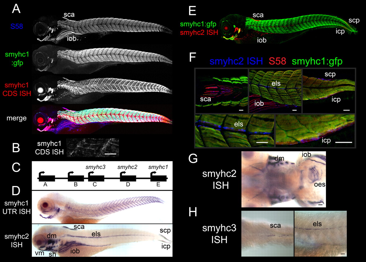
Fig. 1 Different Smyhc genes have distinct expression patterns. (A) At 96 hpf, the smyhc1:gfp reporter gene is expressed in a subset of the slow fibres labelled with the S58 anti slow MyHC Ab or the smyhc1-CDS in situ hybridisation (ISH) probe. The iob is a major muscle that does not express smyhc1:gfp in its slow fibres. (B) High magnification lateral view of smyhc1-CDS in situ hybridisation in posterior trunk showing transcript is concentrated at the ends of the superficial slow fibres. (C) A diagram illustrating the tandem array of Smyhc genes as described by McGuigan et al. (McGuigan et al., 2004) together with the gene names used here. We followed the Bryson-Richardson et al. (Bryson-Richardson et al., 2005) renaming of myhcE as smyhc1 and similarly renamed the adjacent genes smyhc2 and smyhc3. (D) Differential expression of Smyhc genes at 96 hpf, as shown by in situ hybridisation using smyhc1 (1 to 249 bp) or smyhc2 (1 to 324 bp) probes. The sca, els, iob, sh, scp and icp somite-derived muscles, and dorsal and ventral craniofacial muscles express smyhc2. (E) smyhc1:gfp together with smyhc2 (1 to 324 bp) in situ hybridisation at 96 hpf. (F) At 96 hpf, smyhc2 (1 to 324 bp) in situ hybridisation shows colocalisation with slow MyHC S58 antigen but not with smyhc1:gfp in the sca muscle (dorsal view anterior trunk), els or iob muscles (lateral view anterior trunk) or scp or icp muscles (lateral view posterior tail). (G) A deep focus ventral view shows smyhc2 (1 to 324 bp) expression in the oesophagus at 96 hpf. (H) The low sensitivity smyhc3 (1 to 129 bp) in situ hybridisation weakly detects expression in the sca and els muscles at 96 hpf, as shown by dorsal and lateral views of the anterior trunk. Scale bars: 25 μm. Abbreviations: dm, head dorsal muscles; els, embryonic lateralis superficialis; icp, infracarinalis posterior; iob, inferior obliquus; oes, oesophagus; sca, supracarinalis anterior; scp, supracarinalis posterior; sh, sternohyoideus; vm, head ventral muscles. Muscle nomenclature is taken from previous work (Schilling and Kimmel, 1997; Stiassny, 2000; Winterbottom, 1974).