
Fig. 5
- ID
- ZDB-IMAGE-080401-28
- Genes
- Publication
- Nakayama et al., 2008 - Fgf19 is required for zebrafish lens and retina development
- All Figures
- Figures for Nakayama et al., 2008
|
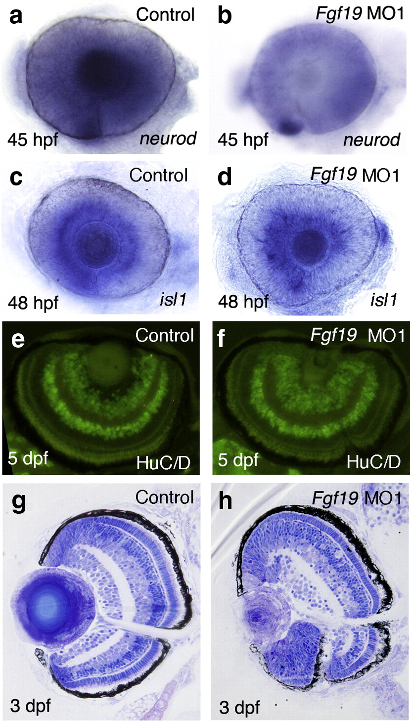
Fig. 5 Neuronal differentiation and lamination of the retina in Fgf19 MO1-injected embryos. (a, b) The expression of neurod in control and Fgf19 MO1-injected embryos at 45 hpf. The expression of neurod in the outer retina was not affected in Fgf19 MO1-injected embryos. Lateral views with anterior to the left and dorsal to the top. (c, d) The expression of isl1 in control and Fgf19 MO1-injected embryos at 48 hpf. The expression of isl1 in the retina was not affected in Fgf19 MO1-injected embryos. Lateral views with anterior to the left and dorsal to the top. (e, f) The detection of HuC/D protein in control and Fgf19 MO1-injected embryos at 5 dpf. HuC/D was normally detected in the amacrine and ganglion cells of Fgf19 MO1-injected embryos. (g, h) Transverse eye sections of control and Fgf19 MO1-injected embryos at 3 dpf. The sections were stained with toluidine blue. The control retina showed a characteristic stratification into three nuclear layers and two plexiform layers. In Fgf19 MO1-injected embryos, the stratification of retinal layers occurred in the dorsal region, although the eye exhibited a reduction in size. However, the lamination of the ventral retina was abnormal in Fgf19 MO1-injected embryos.
Reprinted from Developmental Biology, 313(2), Nakayama, Y., Miyake, A., Nakagawa, Y., Mido, T., Yoshikawa, M., Konishi, M., and Itoh, N., Fgf19 is required for zebrafish lens and retina development, 752-766, Copyright (2008) with permission from Elsevier. Full text @ Dev. Biol.