
Fig. S1
|
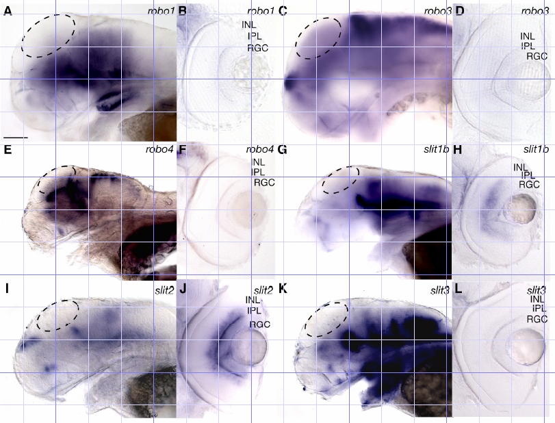
Fig. S1 Expression patterns of slit and robo family members during arborisation and synaptogenesis. Shown are whole-mount lateral views (A, C, E, G, I and K; anterior left, dorsal up) and vibratome sections through the retina (B, D, F, H, J and L; dorsal up) at 76 hpf. robo1 and robo4 are not expressed in the tectum or RGCs (A-B, E-F). robo3 is not expressed widely in the tectum (C) but is expressed in a few discrete tectal cells outside the plane of focus. robo3 is not expressed in RGCs (D). slit1b, slit2, and slit3 are not detectably expressed in the tectum (G, I, K). slit1b is expressed strongly in the inner nuclear layer (H), slit2 is expressed strongly in the inner plexiform layer (J) and slit3 is not expressed in the retina (L). RGC, retinal ganglion cells; IPL, inner plexiform layer; INL, inner nuclear layer. Dashed ovals indicate the tectum. Scale bar = 50μm.