
Fig. 7
- ID
- ZDB-IMAGE-080325-21
- Genes
- Publication
- Hendricks et al., 2008 - Disruption of Esrom and Ryk identifies the roof plate boundary as an intermediate target for commissure formation
- All Figures
- Figures for Hendricks et al., 2008
|
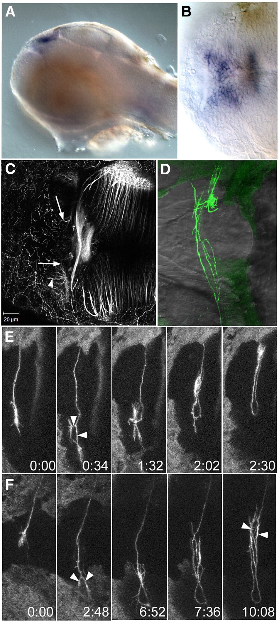
Fig. 7 wnt4a and dnRyk affect commissure formation. Lateral (A) and dorsal (B) view of a 48 hpf embryo, showing expression of wnt4a in the medial habenula. (C) A 3 dpf embryo after injection with a morpholino (MO1) to wnt4a. Habenular afferents have entered the habenula and reached the medial edge (arrows) but have not crossed the roof plate. Axons have innervated the habenula neuropil (arrowhead), which normally occurs only after the commissure has formed. (D) An embryo at 3 dpf after unilateral electroporation with HuC:Gal4/UAS:dnRyk-EGFP at 1 dpf. Abnormal loops and branches are seen within the commissure (n = 15). (E, F) Time series of individual axons show that growth cones reach the contralateral boundary, then bifurcate (arrowheads), turn around and recross the midline (see Supplementary Movies 5 and 6). Time series in (E) was taken without a heated stage, causing slower growth. Time is h:min.
Reprinted from Molecular and cellular neurosciences, 37(2), Hendricks, M., Mathuru, A.S., Wang, H., Silander, O., Kee, M.Z., and Jesuthasan, S., Disruption of Esrom and Ryk identifies the roof plate boundary as an intermediate target for commissure formation, 271-283, Copyright (2008) with permission from Elsevier. Full text @ Mol. Cell Neurosci.