
Fig. 3
- ID
- ZDB-IMAGE-070928-11
- Genes
- Publication
- Kazakova et al., 2006 - A screen for mutations in zebrafish that affect myelin gene expression in Schwann cells and oligodendrocytes
- All Figures
- Figures for Kazakova et al., 2006
|
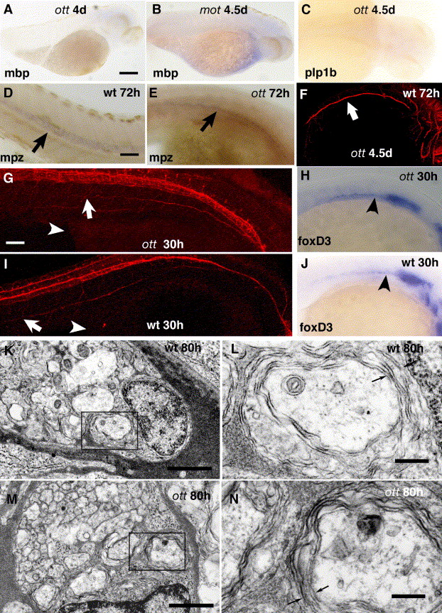
Fig. 3 PLL phenotype of mot/ott mutants. (A–C) Myelin protein expression in ott and mot mutants. Both mutants show a complete lack of mbp and Plp1b expression. (D, E) Mpz expression in the CNS (arrows) of wild type (D) and ott (E) embryos at 72 hpf. Mpz is expressed at reduced levels in ott embryos. (F) Anti-acetylated tubulin staining of the PLL in ott mutants. The axons (arrow) are present but do not extend the whole length of the fish. (G–J) Early development of the PLL nerve and glia precursors. At 30 hpf, axons of the PLL nerve (arrows)of ott mutants (G) have extended less far than in the wild type. Arrowhead—caudal end of yolk sac (I). Prior to myelination, early glial expression (arrowheads) of foxD3 in both mutant (H) and wild type (J) fish. (K–N) Electronmicrographs of transverse sections through the PLL nerve of wild type (K, L) and ott mutant (M, N) fish 80 hpf. The larger diameter axons are ensheathed by loosely packed myelin (arrows) typical of this early stage of development. Scale bars in panels A–C 10 μm; in panels D–F, H and J 5 μm; in panels G and I 3 μm; in panel E 1 μm; in panels K and M; in panels L and N 200 nm.
Reprinted from Developmental Biology, 297(1), Kazakova, N., Li, H., Mora, A., Jessen, K.R., Mirsky, R., Richardson, W.D., and Smith, H.K., A screen for mutations in zebrafish that affect myelin gene expression in Schwann cells and oligodendrocytes, 1-13, Copyright (2006) with permission from Elsevier. Full text @ Dev. Biol.