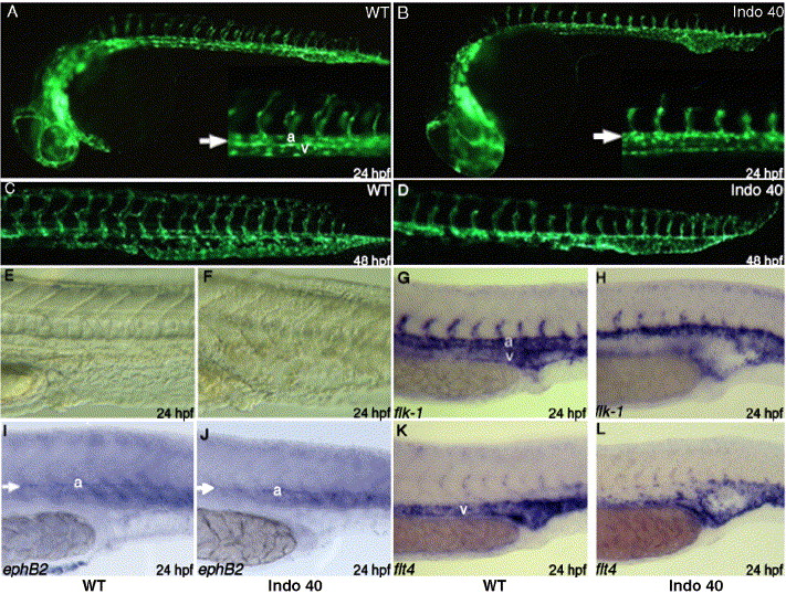
Fig. 3 COX-1 inhibition causes defect in vascular tube formation. Fli1:EGFP transgenic embryos were treated with 40 μM indomethacin at tailbud stage. Compared to untreated embryos (A, C), indomethacin treatment results in distended posterior vasculature at day 1 (B, arrow). Posterior vasculature cannot differentiate into dorsal aorta and posterior cardinal vein (B, inset arrow). At day 2, clear distinction between dorsal aorta and caudal vein can be seen in untreated embryo (C), but a single distended posterior vessel remains in treated embryo (D). In addition, intersomitic vessels are either shortened or absent at day 1 (B, inset) and results in shortened intersomitic vasculature at day 2 (D). Indomethacin treatment results in distention of posterior vasculature at day 1 (F), compared to untreated embryos (E). Staining with flk1, an endothelial marker, reveal the significant reduction or absence of venous staining at day 1 in treated embryos (H), compared to the wild-type (G). While ephB2, arterial marker, staining shows similar staining between wt (I) and treated embryos (J), flt4, vein marker, staining shows absence of veinous staining, especially in the arterial–venous boundary (L).
Reprinted from Developmental Biology, 282(1), Cha, Y.I., Kim, S.H., Solnica-Krezel, L., and Dubois, R.N., Cyclooxygenase-1 signaling is required for vascular tube formation during development, 274-283, Copyright (2005) with permission from Elsevier. Full text @ Dev. Biol.

