
Fig. 4
- ID
- ZDB-IMAGE-070802-6
- Genes
- Publication
- Tessmar-Raible et al., 2007 - Conserved sensory-neurosecretory cell types in annelid and fish forebrain: insights into hypothalamus evolution
- All Figures
- Figures for Tessmar-Raible et al., 2007
|
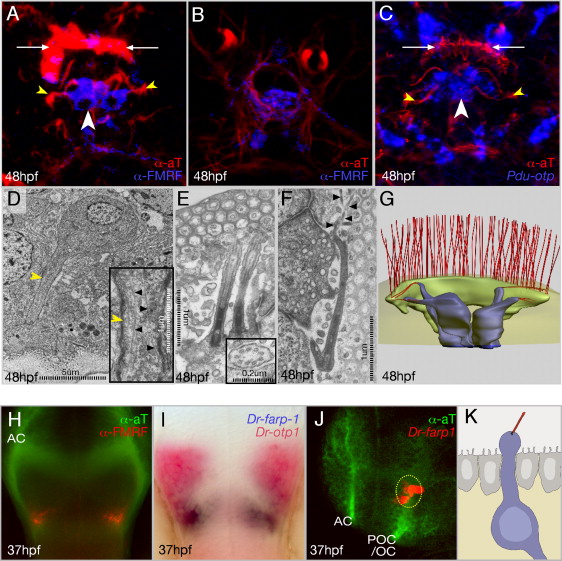
Fig. 4 RFamidergic Neurons in Platynereis and Zebrafish Medial Forebrains. (A–C) Immunostainings/in situ hybridizations of RFamidergic, otp+ cells in the apical plexus of Platynereis embryos. (A and B) Same embryo, different confocal projections, showing the RFamidergic plexus (B) below the contributing cells ([A], same level as [C]). (D–F) Transmission electromicrographs and derived reconstruction (G). (D) TEM view of a medial flask-shaped cell. Inset: enlargement of the dendrite rich in microtubules (black arrowheads). (E) Two cilia protruding from a medial flask-shaped cell to the subcuticular space. Inset: cilium in cross-section. (F) Cilium of the branching type; black arrowheads, ramifications extending underneath the cuticle. (H) zebrafish RFamidergic cells and (I and J) Dr-farp1 transcript in relation to Dr-otp1 (red in [I]) and the axon scaffold (green in [J]). (K) Forebrain CSF-contacting neuron (according to data from Leonhardt, 1980). AC, anterior commissure; POC/ OC, postoptic commissure/optic chiasm. Views: (A–F) apical, (A–C) ventral to the bottom; (G) ventroapical; (H and I) ventral, anterior to the top; (J) lateral, anterior left. White arrows: position of large multiciliated crescent cell as landmark for orientation, yellow arrowheads: microtubule-rich dendrites bearing cilia. Depth of confocal reconstructions: (A and C) 7 μm, (B) 3 μm.
Reprinted from Cell, 129(7), Tessmar-Raible, K., Raible, F., Christodoulou, F., Guy, K., Rembold, M., Hausen, H., and Arendt, D., Conserved sensory-neurosecretory cell types in annelid and fish forebrain: insights into hypothalamus evolution, 1389-1400, Copyright (2007) with permission from Elsevier. Full text @ Cell