
Fig. S2
- ID
- ZDB-IMAGE-070307-65
- Genes
- Publication
- Hans et al., 2007 - Fgf-dependent otic induction requires competence provided by Foxi1 and Dlx3b
- All Figures
- Figures for Hans et al., 2007
|
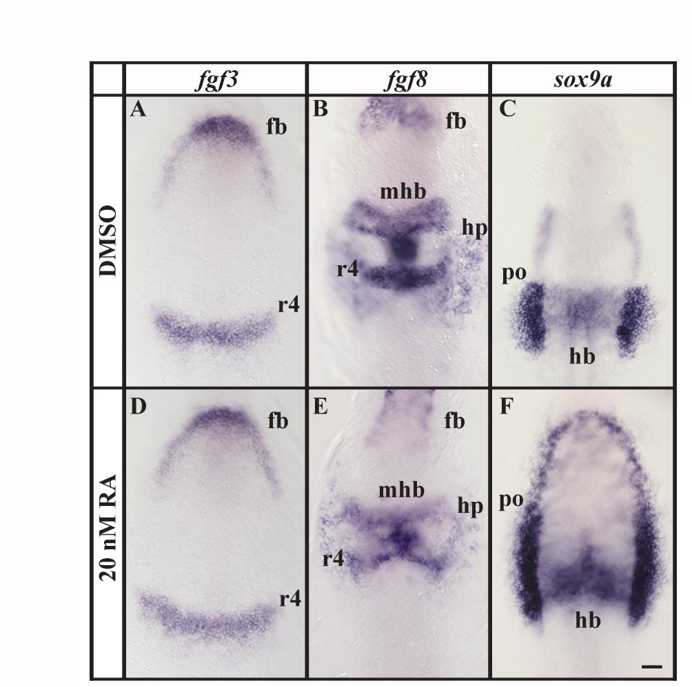
Fig. S2 Retinoic acid treatment has little effect on patterning along the anterior-posterior axis. (A, B, D, E) Expression of fgf3 or fgf8 in embryos treated with 20 nM RA is indistinguishable from control embryos treated with DMSO. (C, F) In RA treated embryos expression of sox9a in the preotic region expands to surround the anterior neural plate border in comparison to control embryos. However, sox9a expression in the hindbrain is identical in RA and DMSO treated embryos. Dorsal views of 1–5-somite stage embryos with anterior towards the top. fb, forebrain; hb, hindbrain; hp, heart primordium; mhb, midbrain-hindbrain border; r4, rhombomere 4; po, preotic region. Scale bar: 40 μm.