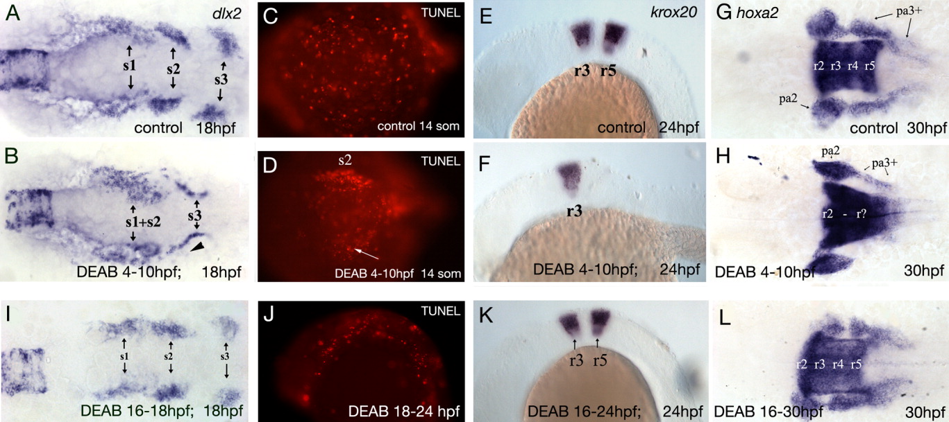
Fig. 4 RA only affects hindbrain specification during gastrulation, likely causing postotic neural crest defects. A: Three streams of neural crest cells are labeled with dlx2 at 18 hpf in untreated embryos as seen from a ventral view. B: Embryos in which RA is blocked during gastrulation show a severe reduction of the postotic neural crest stream. A few postotic neural crest cells (s3) migrate anteriorly and ventrally to the ear and fuse with the second neural crest stream (s2, arrowhead). C: Dorsal view of TUNEL stained 14-som control embryo. Anterior to the left. D: Dorsal view of TUNEL-stained embryo that was treated with DEAB between 4-10 hpf. Dying neural crest cells are present in the second neural crest stream (s2, arrow). E: Expression pattern of krox20 in r3 and r5 and in neural crest cells of pharyngeal arches 2-5 at 24 hpf in untreated embryos. Lateral view. F: No expression was seen in r5 in embryos treated with DEAB during gastrulation. G: In 30-hpf untreated embryos, hoxa2 is expressed in r2-r5 and in neural crest cells of the second and more posterior pharyngeal arches. Ventral view. H: Treatment with DEAB during gastrulation causes an expansion of the posterior hindbrain expression domain of hoxa2 and the posterior boundary is more diffuse. Expression in neural crest cells of pharyngeal arches 3-5 is strongly reduced. Inhibition of RA synthesis post gastrulation (16-30 hpf) does not affect hindbrain patterning or neural crest cell migration. I: In embryos treated with DEAB between 16-18 hpf dlx2 staining reveals that neural crest cells migrate normally in three streams. J: Lateral view of 24-hpf TUNEL-stained embryo that was treated with DEAB between 18-24 hpf. No increase is cell death is observed. Anterior to the left. K: Hindbrain segmentation is not affected if embryos are treated with DEAB post-gastrulation. krox20 (K) and hoxa2 (L) expression in the hindbrain is normal. hoxa2 expression in the posterior pharyngeal arch domain is possibly slightly increased. pa, pharyngeal arch; r, rhombomeres; s1-3, neural crest cell streams 1-3.
Image
Figure Caption
Figure Data
Acknowledgments
This image is the copyrighted work of the attributed author or publisher, and
ZFIN has permission only to display this image to its users.
Additional permissions should be obtained from the applicable author or publisher of the image.
Full text @ Dev. Dyn.

