
Fig. 7
- ID
- ZDB-IMAGE-060920-18
- Genes
- Publication
- Lecaudey et al., 2004 - The zebrafish Iroquois gene iro7 positions the r4/r5 boundary and controls neurogenesis in the rostral hindbrain
- All Figures
- Figures for Lecaudey et al., 2004
|
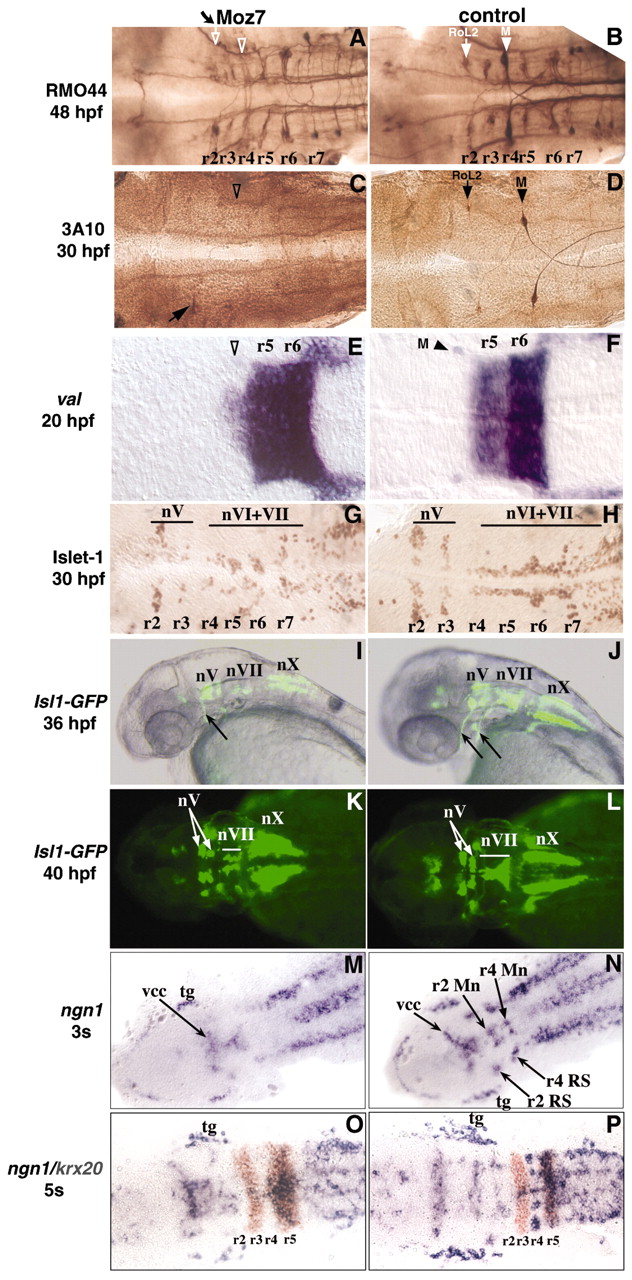
Fig. 7 Knocking-down iro7 results in a reduction of neurogenesis in the anterior hindbrain. (A-D,G,H) Immunohistochemistry using anti-neurofilament RMO44 (A,B), 3A10 (C,D) or anti-Isl1 (G,H) antibodies on Moz7-injected (A,C,G) or control (B,D,H) embryos. (E,F) In situ hybridisation with a val probe on 20 hpf Moz7-injected (E) or control (F) embryos. (A-F) The r4-specific Mauthner neurons (‘M’, arrowhead in B,D,F) and the RoL2 neurons (arrows in B-D) are partially or totally lost after Moz7 injection (empty arrowheads and empty arrows in A,C,E). (I-L) Lateral views (I,J) or dorsal views (K,L) of live Isl1-GFP transgenic embryos injected with Moz7 (I,K) or uninjected (J,L). (G-L) nV, nVI, nVII and nX indicate the motor nuclei of the Vth, VIth, VIIth and Xth nerves, respectively. Arrows in I,J indicate the trigeminal (anterior) and facial (posterior) motor nerves. (M-P) Flat-mounted embryos analysed by in situ hybridisation with the probes indicated on the left (colour-coded) on Moz7-injected (M,O) or Moz7m-injected (N,P) embryos. Anterior is towards the left. Proneural clusters are indicated as follows: Mn, motoneurons; tg, trigeminal ganglion; RS, reticulospinal neurons; vcc, ventrocaudal cluster.