
|
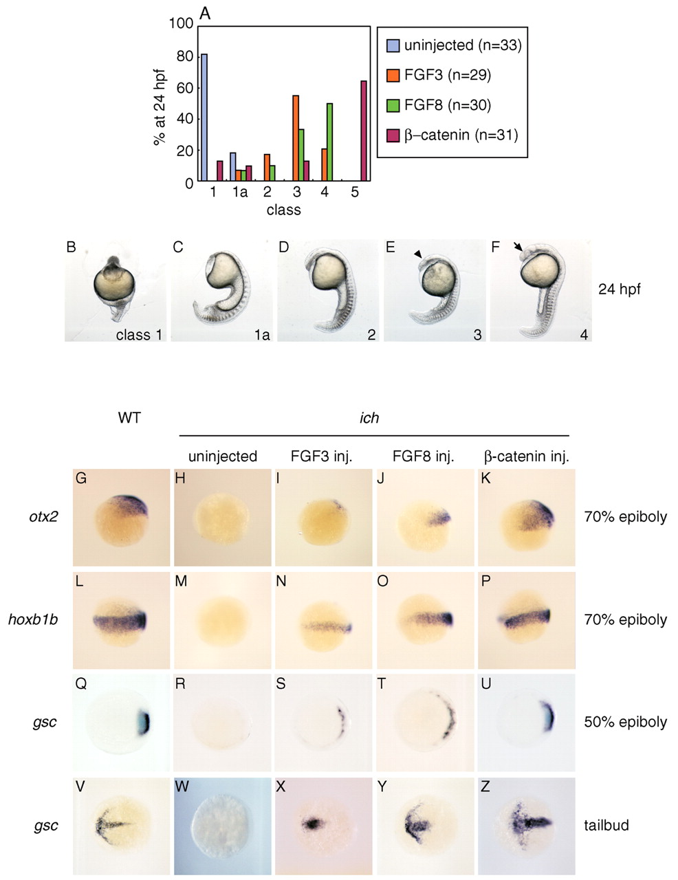
Fig. 1 FGF3 and FGF8 can partially restore AP and DV patterning in ichabod embryos. (A) Partial rescue of ichabod embryos injected with RNAs for FGF3 (orange) or FGF8 (green), compared with uninjected ichabod embryos (blue) or β-catenin-injected embryos (red). (B-F) Phenotypic classes of 24 hpf ichabod embryos (Kelly et al., 2000; Tsang et al., 2004) used for the classification in A and in subsequent figures. Class 1 embryos lack head and trunk; class 1a lack all structures anterior to spinal cord; class 2 lack structures anterior to hindbrain; class 3 form deficient forebrain; class 4 lack notochord (as do the previously mentioned classes) but have a complete AP axis. Only class 5 embryos (not shown) appear normal at 24 hpf. The arrowhead in E indicates the position of the midbrain/hindbrain boundary; the arrow in F points to aneye. (G-Z) FGFs restore expression of markers of anterior and posterior neurectoderm and organizer. FGF3 and FGF8 induce otx2 (H-J), hoxb1b (M-O) and gsc (R-T,W-Y). Expression is compared to wild-type (G,L,Q,V), uninjected ichabod (H,M,R,W) and ichabod embryos injected with β-catenin RNA (K,P,U,Z). Embryos are shown at 70% epiboly (G-P, lateral views), 50% epiboly (Q-U, animal pole views) and 10 hpf tailbud stage (V-Z, dorsal views).