
|
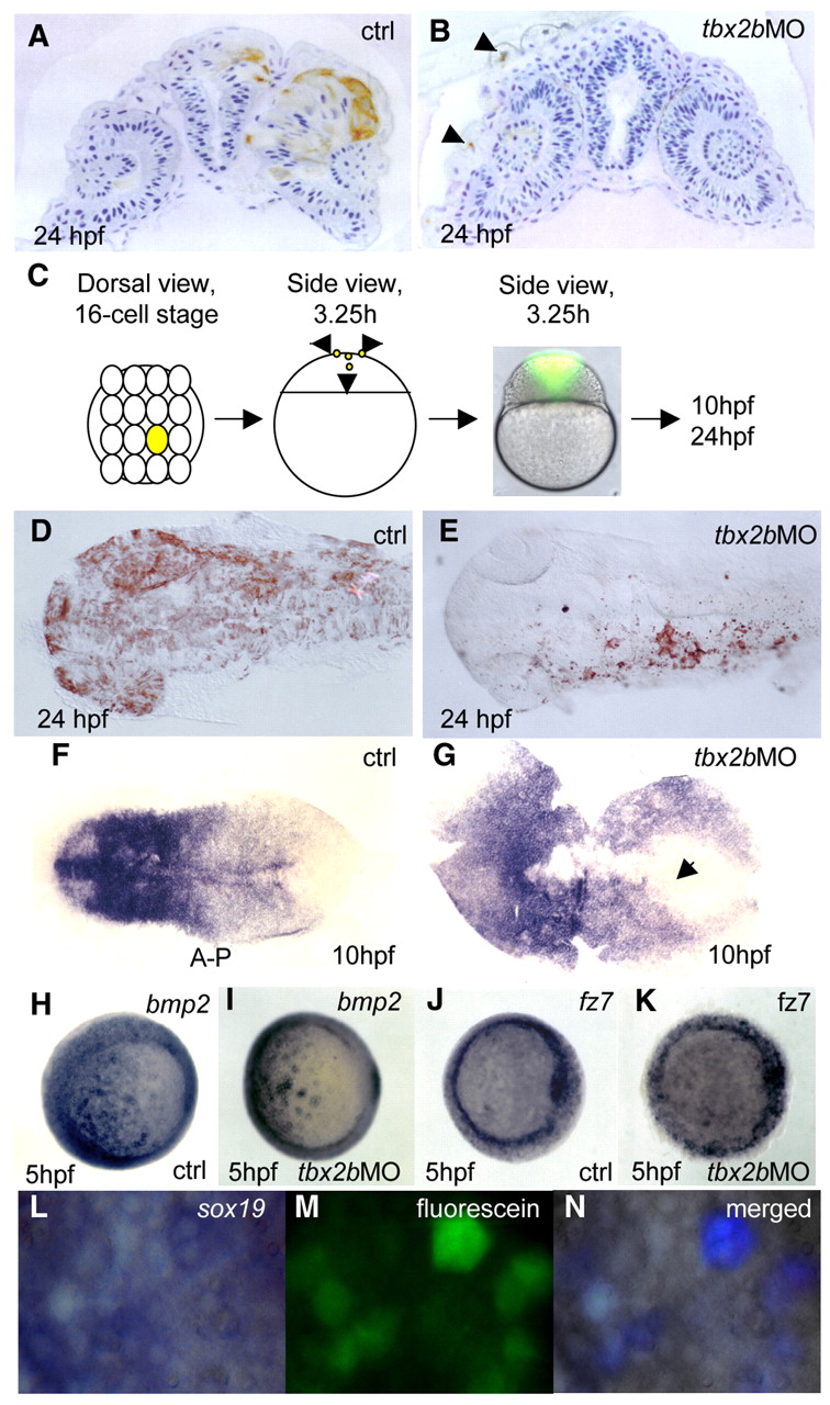
Fig. 2 Tbx2b is required for cells to adopt a neural fate. Deep cells from embryos co-injected with fluorescein-dextran (70 kDa) and tbx2b MO at 30% epiboly were transplanted into homochronic wild-type hosts. (A) Cross-section through the neuroretina and brain shows transplanted control labelled cells in the CNS. By contrast (B) transplanted Tbx2b-deficient cells were present only in the epidermis (arrowheads). (C) One central blastomere of 16-cell stage embryos was injected with fluorescein either with or without tbx2b MO, and embryos were allowed to develop to 24 hpf. Both control and tbx2b morphants developed normally and showed a similar distribution of labelled clones in vivo at 3.25 hours. (D) Dorsal view of 24 hpf control embryo shows labelled cells in neural plate and epidermis. (E) tbx2b morphant labelled cells were present only in epidermis. (F,G) Whole-mount in situ hybridization with the pan-neural marker sox19 shows a delay in convergence of the neural plate in tbx2b morphant (arrow in G) compared with control (F). (H-K) Whole-mount in situ hybridization with bmp2 (H,I) and fz7 (J,K) show similar expression patterns in control and Tbx2b morphant at 5 hpf. (L-N) Transplanted fluorescein labelled Tbx2b-deficient cells continue to express sox19 in 5 hpf wild-type host. (N) The merged photographs were enhanced in Adobe Photoshop such that only the simultaneous presence of blue (whole-mount in situ hybridization signal) and green (fluorescence) produced a purple signature. Otherwise, cells remain grey.