
Fig. 4
- ID
- ZDB-IMAGE-050518-1
- Genes
- Publication
- Vihtelic et al., 2005 - Lens opacity and photoreceptor degeneration in the zebrafish lens opaque mutant
- All Figures
- Figures for Vihtelic et al., 2005
|
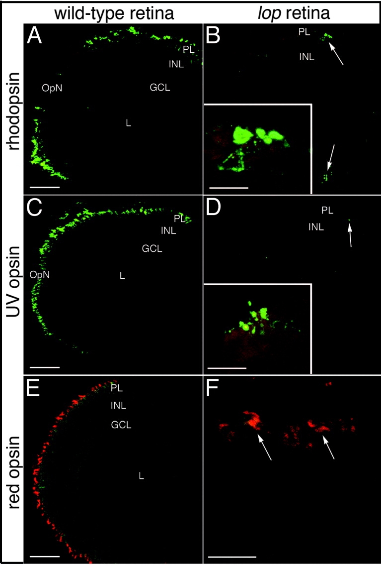
Fig. 4 Rod and cone opsin immunolocalization in wild-type and lop mutant retinas. A,C,E: Immunolocalization of rhodopsin (rods), ultraviolet (UV) opsin (short single cones), and red opsin (double cone cell member), respectively, in wild-type frozen retinal sections. B,D,F: The corresponding localization patterns in the mutant retinas. The rhodopsin and UV opsin proteins are restricted to the retinal margins of the mutants (B and D, respectively; arrows). The insets show higher magnification images of the few remaining opsin-expressing cells located at the retinal margins. Red opsin expression in the mutant retinas was also limited to the retinal marginal zone. E,F: The wild-type red opsin expression pattern and a high magnification image of the red opsin-expressing cells at the mutant retinal margin (arrows) are shown in E and F, respectively. PL, photoreceptor layer; INL, inner nuclear layer; GCL, ganglion cell layer; OpN, optic nerve; L, lens. Scale bars 50 μm in A,C,E, 20 μm in B,D,F.