
Figure 5
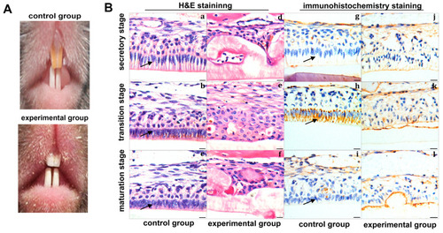
Correlation between fluoride and ClC-3 in DF mouse model. (A) Appearance of mouse incisors in control (upper lane) and DF mouse model (lower lane). (B) Histological changes in mouse incisors and the related ClC-3 changes. (a~f) H&E staining. Images a~f illustrate sequential stages of mouse mandibular incisor enamel differentiation. In the secretory stage, normal ameloblasts are tall, columnar and regularly aligned (a); they shorten during the transition (b) and maturation stages (c). In DF-treated samples, ameloblasts are irregularly arranged, cysts are present, and the enamel is malformed (d~f). (g~l): sABC immunohistochemical staining for CLC-3 in tooth. Images (h) shows strong positive staining for CLC-3 in ameloblasts at the transition stage in the control group. By contrast, positive staining for CLC-3 is observed in DF-treated ameloblasts at all three stages (j~l). Black arrows point to the ameloblasts. Scale bars = 10 μm. |