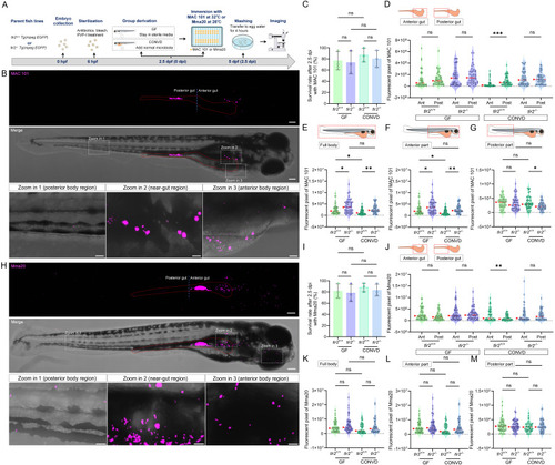
TLR2 and the microbiome modulate bacterial gut distribution and dissemination after immersion infection with MAC 101 and Mma20. (A) Schematic of the experimental workflow. Germ-free (GF) and conventionalized (CONVD) zebrafish larvae derived from tlr2 wild-type and mutant lines were immersed in egg water containing either mCherry-labeled M. avium complex (MAC 101) or DsRed-labeled M. marinum (Mma20) bacteria for 2.5 days. (B) Representative fluorescence images showing MAC 101 localization primarily in the gut, with dissemination to anterior and posterior regions (representative images are tlr2 wild-type larvae under the germ-free (GF) condition). Bacteria are shown in magenta. Scale bar: 100 µm for full-body images; 25 µm for zooms. (C) Survival rates for all groups after MAC 101 infection showing no significant differences between genotypes or microbial conditions. The results are based on 3 independent experiments. Error bars represent mean ± SD. (D) Distribution of MAC 101 within the gut shows preferential localization to the posterior gut in wild-type CONVD larvae, whereas this regional bias is lost in tlr2 mutants and GF conditions. (E-F) Quantification of full body (E) and anterior region (F) bacterial burden (fluorescent pixel count) reveals significantly higher MAC 101 load in tlr2 mutants compared to the wild type under both GF and CONVD conditions. (G) In the posterior body region, significantly more MAC 101 was detected in tlr2 wild-type larvae than in mutants under CONVD conditions, but not under GF conditions. For MAC 101 infection, the data from GF tlr2+/+ (n=46) group, GF tlr2-/- (n=44) group, CONVD tlr2+/+ (n=52) group and CONVD tlr2-/- (n=48) group are based on three independent experiments. (H) Representative fluorescence images showing Mma20 localization primarily in the gut, with dissemination to anterior and posterior regions. (I) Survival rates for all groups after Mma20 infection showing no significant differences between genotypes or microbial conditions. The results are based on 3 independent experiments. Error bars represent mean ± SD. (J) Distribution of Mma20 in the gut shows higher abundance in the anterior gut in wild-type CONVD larvae, opposite to the distribution pattern observed with MAC 101. (K-M) Quantification of Mma20 bacterial burden in the full body (K) and anterior region (L) showing a non-significant trend toward higher levels in tlr2 mutant larvae. (M) No significant differences in Mma20 burden were detected in the posterior region among groups. For Mma20 infection, the data from GF tlr2+/+ (n=50) group, GF tlr2-/- (n=51) group, CONVD tlr2+/+ (n=45) group and CONVD tlr2-/- (n=50) group are based on three independent experiments. Statistical significant difference was determined by one-way ANOVA, red arrows point to the median, ns, non-significant, *, P < 0.05, **, P < 0.01, ***, P < 0.001.
|
