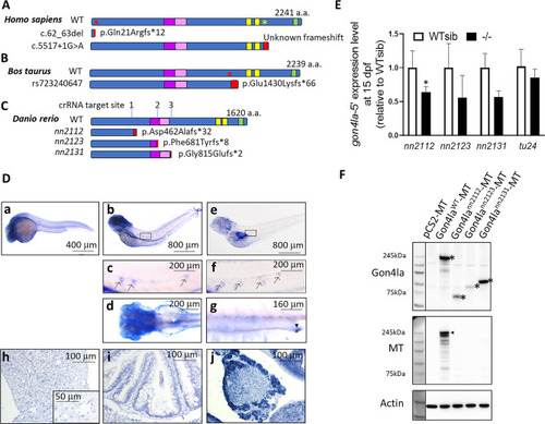
Evolutionary conservation, generation of mutant alleles, and spatiotemporal expression of zebrafish gon4la. (A–C) Schematic comparison of GON4L protein domains and clinical/natural variants in humans, cattle, and zebrafish. (A) WT and patient GON4L protein (predicted by SpliceAI). Red asterisk: premature stop codon resulted from a 2-bp deletion (c. 62_63del); yellow asterisk: canonical splice variant (c.5517+1G>A). Both variants were predicted to be subject to nonsense mediated RNA decay6. The c.5517+1G>A variant is predicted to disrupt splicing (e.g., leading to intron retention and frame shift of protein product). Due to lack of information, the abnormal transcript(s) in patients carrying c.5517+1G>A variant require experimental confirmation. (B) Wild type and predicted mutant cattle GON4L proteins. Red asterisk indicates premature stop codon resulting from the 1-bp deletion. (C) Zebrafish Gon4la protein sequences, showing three CRISPR/Cas9-targeted indels resulting in frameshift mutations and truncated proteins. Important domains are as indicated: purple box: N-terminal YY1-binding domain (N-YBD); pink box: putative transactivation domain (TD); yellow box: PAH domain; green box: SANT domain. Red box: additional peptide tail due to frameshift caused by introduced indels. (D, a–g) Spatiotemporal expression of gon4la mRNA via whole-mount in situ hybridization. (a) gon4la is strongly expressed in the brain at 1 dpf. At 3 (b, c) and 5 (e, f) dpf, gon4la is also detected in the neuromasts of the lateral lines (black arrows). Additionally, gon4la expression is observed in the gut looping region (d, dorsal view, white arrowhead) at 3 dpf and in the posterior gut and cloaca at 5 dpf (g, lateral view, black arrowhead). (D, h–j) Section in situ hybridization analysis of gon4la in adult tissues. (h) In the liver, gon4la is expressed in hepatocytes and sinusoidal endothelial cells (inset). (i) In the gut, gon4la is expressed in intestinal epithelial cells. (j) In the pancreas, gon4la is weakly expressed in islet cells and strongly expressed in exocrine cells. (E) qRT-PCR analysis of gon4la mRNA expression in WT siblings and gon4la homozygous mutants at 15 dpf. For comparison, gon4lbtu24 WT siblings and homozygous mutants at 5 dpf larvae were genotyped and analyzed. *, p < 0.05; n = 6. (F) Protein products expressed from WT and mutant gon4la constructs were detected by Western blot. Full-length Gon4la is detected in WT samples (asterisk: N-terminal; arrowhead: C-terminal Myc-tag, denoted as MT). Mutant alleles produce truncated proteins lacking the Myc tag. Actin was used as loading controls.
|
