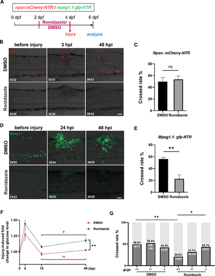
Regeneration defects caused by macrophage depletion can be rescued by the gcga mutation. (A) Schematic of the Ronidazole treatment workflow. (B and D) Representative images of Tg(npsn: mCherry-NTR) (B) and Tg(mpeg1.1: GFP-NTR) (D) after injury following DMSO or Ronidazole treatment. Scale bar = 50 µm. (C,E) Quantification of the proportion of larvae with axons successfully crossing the injury site following neutrophil (C) and macrophage (E) depletion. Error bars represent means ± s.d. Data were analysed by paired t‐test. (F) Relative glucose levels in the injured group compared to the uninjured group in macrophage-depleted and control larvae at 4, 16 and 48 hpi. Data were analysed by two-way ANOVA. ** The statistically significant difference in injury-induced glucose changes between Ronidazole-treated larvae and DMSO controls at 48 hpi. # The statistically significant difference in glucose changes between 16 and 48 hpi within the Ronidazole-treated group. ns indicates no statistically significant difference between 16 and 48 hpi in the DMSO-treated group. This graph combines data from three biological replicates. (G) Quantification of the proportion of larvae with axons successfully crossing the injury site following macrophage depletion in gcga siblings and mutants. Data were analysed by the chi-square test. ns, p > 0.05; **p < 0.01; *p < 0.05; #p < 0.05.
|
