FIGURE

Figure 5

ID
ZDB-FIG-260316-83
Publication
Wang et al., 2026 - Zebrafish PRL-3 Regulates Yolk Syncytial Layer Integrity and Actomyosin Contractility During Epiboly
Other Figures
All Figure Page
Back to All Figure Page
Figure 5

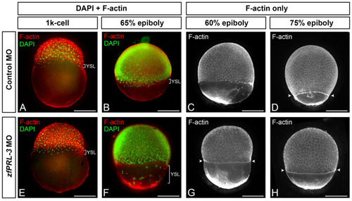
Whole-mount F-actin and nuclear staining reveals abnormal YSL distribution and premature actomyosin ring formation in zfPRL-3 morphants. Representative images of embryos injected with 4ng control morpholino (MO) or zfPRL-3 MO and stained for F-actin (phalloidin) and nuclei (DAPI; 4′,6-diamidino-2-phenylindole). Panels are grouped by staining modality to highlight different readouts: nuclei (DAPI) together with F-actin to provide positional context for YSL nuclei, versus F-actin–only staining to better resolve the actomyosin ring. Brackets in panels (A,B,E,F) indicate the region corresponding to the yolk syncytial layer (YSL). Arrowheads in panels (D,G,H) indicate the actomyosin ring. (AD) Control morphants. (EH) zfPRL-3 morphants. In zfPRL-3 morphants at ~60% epiboly, bracket in panel (F) highlight dispersed external YSL-associated nuclei, and arrowheads in panel (G) indicate the early appearance of the actomyosin ring relative to controls. Scale bars = 250 μm.

Expression Data

Expression Detail
Antibody Labeling
Phenotype Data
Fish:
Knockdown Reagent:
Observed In:
Stage Range: 1k-cell to 75%-epiboly

Phenotype Detail
Acknowledgments
This image is the copyrighted work of the attributed author or publisher, and ZFIN has permission only to display this image to its users. Additional permissions should be obtained from the applicable author or publisher of the image. Full text @ Int. J. Mol. Sci.