FIGURE

Fig. 5

ID
ZDB-FIG-260311-87
Publication
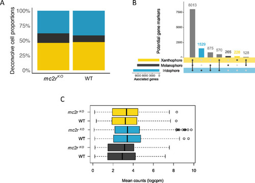
Barreiro-Docío et al., 2026 - Loss-of-function mutations in the melanocortin-2-receptor (mc2r) lead to skin hyperpigmentation in teleost fish
Other Figures
All Figure Page
Back to All Figure Page
Fig. 5

Pigment cell distribution and differential gene expression in mc2rKO mutant and WT zebrafish skin. (A) Estimation of the proportion of pigment cells in the skin of mc2rKO and WT fish via deconvolution analysis of bulk transcriptome data, with single-cell data used as a reference. (B) UpSet plot demonstrating significantly background-dependent differentially expressed genes (DEGs) from skin single-cell transcriptome data63 for xanthophore, melanophore, and iridophore cells. Distinctively expressed genes for iridophores, melanophores, and xanthophores are indicated with blue, black, and yellow bars, respectively. (C) Box plot of the counts of pigment cell types in WT and mc2rKO skin, based on DEGs unique to each pigment cell type, as identified in the UpSet plot. The black lines represent the median, the whiskers indicate the 25th and 75th percentiles, and the points represent outliers. Melanophores are represented in black, xanthophores in yellow, and iridophores in blue.

Expression Data

Expression Detail
Antibody Labeling
Phenotype Data

Phenotype Detail
Acknowledgments
This image is the copyrighted work of the attributed author or publisher, and ZFIN has permission only to display this image to its users. Additional permissions should be obtained from the applicable author or publisher of the image. Full text @ Sci. Rep.