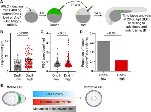
Control of motility loss by dnd1. (A) iPGCs were transplanted either into PGC-depleted (experiments presented in B,C) or into wild-type (experiment presented in D and Fig. S5B) hosts. At 28 hpf, transplanted iPGCs were identified by the fluorescent marker (green). Endogenous PGCs in wild-type hosts are marked in orange. (B,C) iPGC displacement (B) and speed (C). Unpaired two-tailed t-test. Error bars: s.e.m. 91 (Dnd1-low) and 83 (Dnd1-high) PGCs. (D) iPGCs were transplanted into wild-type embryos that were raised and outcrossed. Fish were considered positive if they produced fluorescently labeled embryos. P=0.033, odds ratio=3.62 (Fisher's exact test); 41 (Dnd1-low) and 44 (Dnd1-high) animals. (E) Motile PGCs (left) express high levels of Dnd1 and are polarized [Actin in green at the cell front, and back markers at the rear (magenta)]. Later, dnd1 level declines (red gradient), motility is reduced (blue gradient), while tissue rigidity (e.g. ECM level) and adhesion rise (gray gradient) culminating in stop of migration.
|
