IMAGE

Fig. 4.

ID
ZDB-IMAGE-260311-66
Publication
Tarbashevich et al., 2026 - Cell-autonomous control coupled with tissue context regulates the cessation of migration at the site of organ development
All Figures
Figures for Tarbashevich et al., 2026
Image
Figure Caption

Fig. 4.

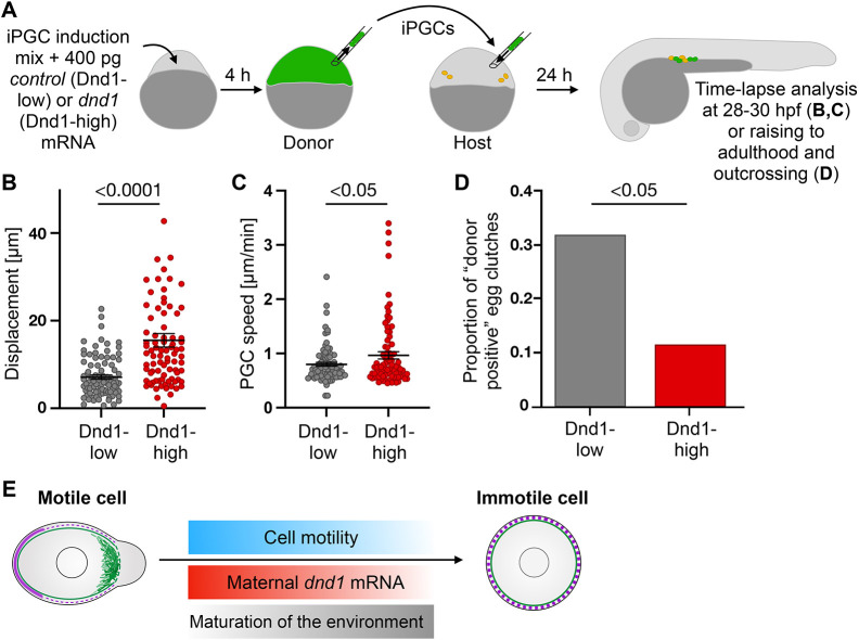
Control of motility loss by dnd1. (A) iPGCs were transplanted either into PGC-depleted (experiments presented in B,C) or into wild-type (experiment presented in D and Fig. S5B) hosts. At 28 hpf, transplanted iPGCs were identified by the fluorescent marker (green). Endogenous PGCs in wild-type hosts are marked in orange. (B,C) iPGC displacement (B) and speed (C). Unpaired two-tailed t-test. Error bars: s.e.m. 91 (Dnd1-low) and 83 (Dnd1-high) PGCs. (D) iPGCs were transplanted into wild-type embryos that were raised and outcrossed. Fish were considered positive if they produced fluorescently labeled embryos. P=0.033, odds ratio=3.62 (Fisher's exact test); 41 (Dnd1-low) and 44 (Dnd1-high) animals. (E) Motile PGCs (left) express high levels of Dnd1 and are polarized [Actin in green at the cell front, and back markers at the rear (magenta)]. Later, dnd1 level declines (red gradient), motility is reduced (blue gradient), while tissue rigidity (e.g. ECM level) and adhesion rise (gray gradient) culminating in stop of migration.

Acknowledgments
This image is the copyrighted work of the attributed author or publisher, and ZFIN has permission only to display this image to its users. Additional permissions should be obtained from the applicable author or publisher of the image. Full text @ Development