FIGURE

Fig. 1.

ID
ZDB-FIG-260311-47
Publication
Tarbashevich et al., 2026 - Cell-autonomous control coupled with tissue context regulates the cessation of migration at the site of organ development
Other Figures
All Figure Page
Back to All Figure Page
Fig. 1.

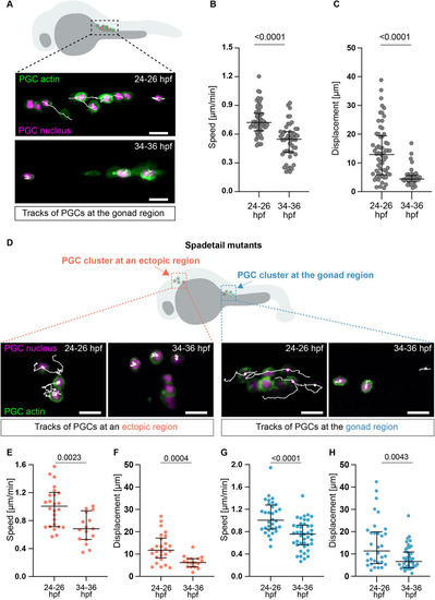
Reduction in PGC migration speed during development. (A) PGCs at the gonad region at 24-36 hpf. (B,C) PGC speed (B) and displacement (C) over 60 min (24-26 hpf: 8 embryos, 60 PGCs; 34-36 hpf: 12 embryos, 48 PGCs). (D) PGC clusters at the gonad and the head at 24-36 hpf. (E,F) PGC speed (E) and displacement (F) in ectopic clusters over 60 min (24-26 hpf: 11 embryos, 26 PGCs; 34-36 hpf: 10 embryos, 17 PGCs). (G,H) PGC speed (G) and displacement (H) at the gonad over 60 min (24-26 hpf: 12 embryos, 32 PGCs; 34-36 hpf: 11 embryos, 42 PGCs). Unpaired two-tailed t-test. Data are median ± error bars (IQR). Scale bars: 20 µm.

Expression Data

Expression Detail
Antibody Labeling
Phenotype Data
Fish:
Observed In:
Stage Range: Prim-5 to Prim-25

Phenotype Detail
Acknowledgments
This image is the copyrighted work of the attributed author or publisher, and ZFIN has permission only to display this image to its users. Additional permissions should be obtained from the applicable author or publisher of the image. Full text @ Development