FIGURE

Figure 4.

ID
ZDB-FIG-260311-459
Publication
Surl et al., 2026 - A Dosage-Dependent Dominant-Negative Mechanism of SLC45A2(W74R) in Autosomal Dominant Oculocutaneous Albinism Revealed by Zebrafish Modeling
Other Figures
All Figure Page
Back to All Figure Page
Figure 4.

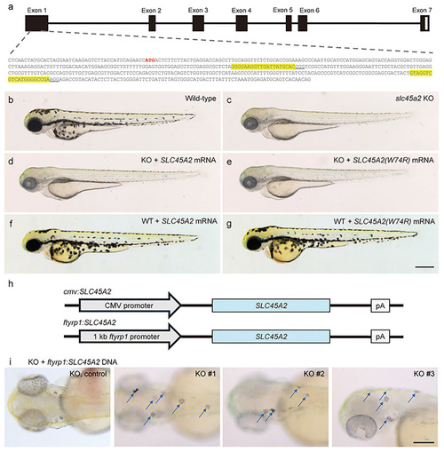
Establishment of slc45a2 KO zebrafish and effects of overexpression of SLC45A2 or SLC45A2(W74R) mRNA in KO zebrafish. (a) Schematic view of the zebrafish slc45a2 gene structure and CRISPR target site in exon 1 (arrow). Predicted protein structure resulted from the frameshift mutation in slc45a2 KO zebrafish. Multiple transmembrane domains in the wild-type (WT) Slc45a2 protein are marked in gray. (b, e) Wild-type zebrafish at 52 hpf (n = 20). (c) Uninjected control KO zebrafish (n = 20). (d) SLC45A2 mRNA-injected KO zebrafish (n = 20). (f) SLC45A2 mRNA-injected WT zebrafish (n = 20). (g) SLC45A2(W74R) mRNA-injected WT zebrafish (n = 20). (h) Schematic view of SLC45A2 expression vectors under control of the cmv promoter or melanocyte-specific ftyrp1 promoter. (i) Detection of rescued melanocytes (arrows) in representative individual KO zebrafish (KO 1–3, n = 15; KO control, n = 4) at 72 hpf after injection with ftyrp1:SLC45A2 plasmid DNA. Five to seven positive melanocytes were detected in an individual KO zebrafish. Scale bar: 400 µm.

Expression Data

Expression Detail
Antibody Labeling
Phenotype Data
Fish:
Observed In:
Stage: Long-pec

Phenotype Detail
Acknowledgments
This image is the copyrighted work of the attributed author or publisher, and ZFIN has permission only to display this image to its users. Additional permissions should be obtained from the applicable author or publisher of the image. Full text @ Transl Vis Sci Technol