FIGURE

FIGURE 4

ID
ZDB-FIG-260123-36
Publication
Christiansen et al., 2026 - Innervation Drives Postembryonic Expansion of the Zebrafish Anterior Lateral Line System
Other Figures
All Figure Page
Back to All Figure Page
FIGURE 4

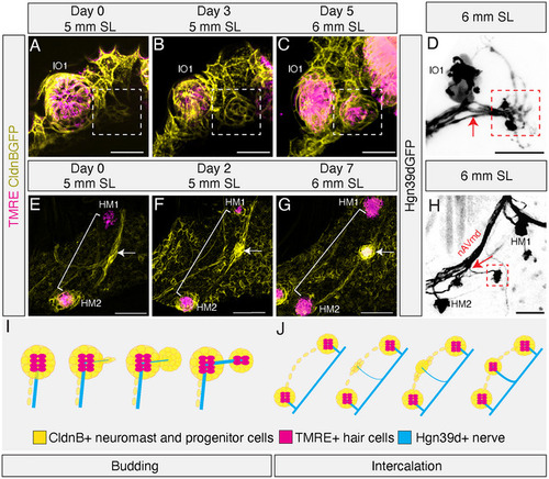
Development of anterior lateral line superficial neuromasts by budding and intercalation mechanisms. (A–C) High magnification confocal time series of lateral line tissue (CldnB:GFP; yellow) and mature neuromasts (TMRE; magenta) showing the budding mechanism of neuromast formation in a single live specimen from 5–6 mm SL. (A) At Day 0, canal neuromast IO1 extends a posterior projection of a few CldnB: GFP‐labeled cells (white box). (B) By Day 3, the cells from the posterior projection have proliferated to form a protoneuromast (white box). (C) By Day 5, this tissue has differentiated into a mature daughter neuromast expressing TMRE (white box) lying directly posterior to the founder neuromast IO1. (D) Confocal image of a live Tg(Hgn39D:GFP) 6 mm SL specimen showing a direct nerve branch (red arrow) extending from the ganglion, which also innervates the founder neuromast (IO1), to the daughter (red box) neuromast. (E–G) High magnification confocal time series of the lateral line tissue (CldnB:GFP; yellow) and mature neuromasts (TMRE; magenta) showing the intercalation mechanism of neuromast formation in a single live specimen from 5–6 mm SL. These images were manually segmented to remove unwanted signal from superficial epithelial tissue. (E) At Day 0, a HM superficial neuromast is beginning to form (white arrow) from a thickening of interneuromast tissue (white bracket) between two mature canal neuromasts, HM1 and HM2. (F) At Day 2, the interneuromast tissue has proliferated into a rosette shape (white arrow). (G) By Day 7, the interneuromast tissue has differentiated into a mature superficial neuromast with a TMRE signal (white arrow). (H) Confocal image of a live Tg(Hgn39D:GFP) 6 mm SL specimen showing a new nerve branch (red arrow) extending from the main HM nerve between the HM1 and HM2 branches to innervate the newly formed HM superficial neuromast (red box). (I–J) Schematics of neuromast formation processes: CldnB:GFP‐labeled neuromast and progenitor cells (yellow), TMRE+ hair cells (magenta), and Hgn39d+ nerves (cyan). (I) Schematic of the budding process. (J) Schematic of the intercalation process.

Expression Data

Expression Detail
Antibody Labeling
Phenotype Data

Phenotype Detail
Acknowledgments
This image is the copyrighted work of the attributed author or publisher, and ZFIN has permission only to display this image to its users. Additional permissions should be obtained from the applicable author or publisher of the image. Full text @ J. Comp. Neurol.