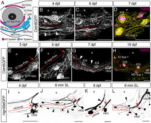
Mechanisms of larval superficial neuromast formation from late‐forming primordia. (A) Schematic of CldnB‐labeled lateral line primordia derived from AD (magenta) and AV (cyan) placodes. (B–H) Confocal images of live Tg(CldnB:GFP) specimens. (B–D) Development of the mandibular superficial line from its migrating primordium (MD Sup Prim) at (B) 4 dpf; (C) 6 dpf; and (D) 7 dpf. (B) The mandibular superficial primordium (red bracket) extends from HM2 by 4 dpf, parallel to the mandibular primordium (white arrowheads). (C) By 6 dpf the primordium has begun to condense into a protoneuromast (red box). (D) At 7 dpf, the first neuromast of the MD superficial line is in place (MD Sup 1; red box); CldnB:GFP (yellow); TMRE (magenta) labels hair cell mitochondria within the neuromast. (E–H) Development of the infraorbital superficial line from its migrating primordium (IO Sup Prim). Extension of the IO superficial primordium (red bracket) adjacent to HM1 at (E) 3 dpf; (F) 5 dpf; (G) 7 dpf; and (H) 10 dpf; where it condenses into the first neuromast of the IO superficial line (IO Sup 1; red box). (E) The infraorbital superficial primordium originates adjacent to HM1 (red arrow) and extends a few cells ventrally. (F) At 4 dpf, the primordium has extended past HM2, running parallel to the IO canal line. (G) At 7 dpf, the primordium has extended parallel to the IO canal line (white arrowheads) to its terminal location adjacent to the eye between IO1 and IO2 (red box), where the terminus has condensed into a protoneuromast. (H) At 10 dpf, this terminus (red box) has differentiated into the first neuromast in the IO superficial line, labeled with CldnB:GFP (yellow) and neuromast hair cell marker TMRE (magenta). (I–H) Confocal images of live (I, K) and fixed (J, H) Tg(Hgn39D:GFP) specimens immunolabeled for GFP. (I) The nAVmds nerve (red bracket) is shown at 4 dpf, branching from gAV near HM2 (red arrow), and running parallel to the MD canal nerve (black arrowhead). (J) At 6 mm SL, the nAVmds nerve (red arrowheads) has extended anteriorly to innervate neuromasts in the mandibular superficial line (red boxes), continuing to run parallel to the MD canal nerve (black arrowheads). (K) The nAVios nerve (red bracket) branches off from gAV near HM1 (red arrow) and runs parallel to the IO canal nerve (black arrowhead). (L) At 6 mm SL, the nAVios nerve (red arrow) has formed a ventral branch, with both the original and ventral branches (red brackets) running parallel to the IO canal nerve (black arrowhead) and innervating neuromasts in the infraorbital superficial line (red boxes). e, eye.
|
