
Fig. 4
- ID
- ZDB-FIG-260114-19
- Publication
- Jia et al., 2025 - Swimming motions evoke Piezo1-dependent Ca2+ events in vascular endothelial cells of larval zebrafish
- Other Figures
- All Figure Page
- Back to All Figure Page
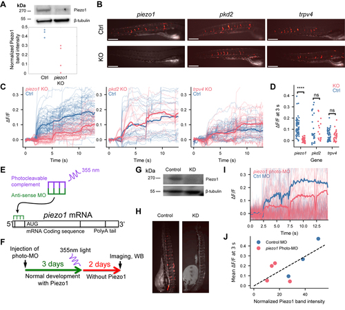
Piezo1 channels are required for motion-evoked EC Ca2+ events.(A) western blot quantification of Piezo1 expression in whole-animal lysates from control and Piezo1 gRNA-injected fish (5 dpf), normalized to β-tubulin band intensity. (B) Relative GCaMP5G fluorescence 3 s after electrical field stimulus in knockout (piezo1, pkd2, or trpv4) versus control Tg(kdrl:Gal4;UAS:GCaMP5G) fish. Scale bars 500 μm. (C) ΔF/F traces for individual blood vessel segments. Electric field stimuli at 2, 6, and 11 s. Bold lines indicate mean over all measured vessels in each group of fish. (D) ΔF/F 3 seconds after the first electrical field stimulus. (C – D) n = 6 controls, n = 3 KO piezo1; n = 3 controls, n = 3 KO pkd2; n = 4 controls, n = 3 KO trpv4. (E) Photo-morpholino (MO) mechanism of action. (F) Experimental design for temporally restricted photo-morpholino knockdown of Piezo1. (G) Western blot measuring Piezo1 knockdown by photo-MO. (H) Relative GCaMP5G fluorescence 3 s after electrical field stimulus in scrambled and piezo1 photo-MO treated fish. (I) ΔF/F traces for individual blood vessel segments. Bold line represents the mean over all measured vessels. (J) Mean GCaMP5G ΔF/F over intersegmental vessels 3 s after electrical field stimulus compared to post-hoc western blot Piezo1 band intensity normalized to β-tubulin band intensity (Pearson R2 = 0.49). (I, J) Control morpholino: 34 vessels over n = 3 fish, piezo1 photo-morpholino 68 vessels over n = 4 fish. Statistical tests: (D) Mixed-effects linear model considering individual fish and CRISPR/Cas9 treatment of each vessel, piezo1: z = −4.8, p = 1.4e-6; pkd2: z = 0.48, p = 0.63; trpv4: z = 0.002, p = 0.998. Additional supporting data in Figure S4. |