
Fig. 2
- ID
- ZDB-FIG-260114-17
- Publication
- Jia et al., 2025 - Swimming motions evoke Piezo1-dependent Ca2+ events in vascular endothelial cells of larval zebrafish
- Other Figures
- All Figure Page
- Back to All Figure Page
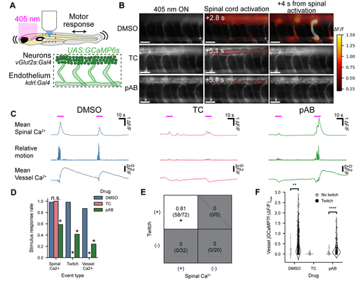
Calcium events in ECs require muscle contraction. (A) Experimental setup. Violet light on the eyes evoked escape attempts; Ca2+ responses were monitored in the spinal cord and in vascular endothelial cells. (B) Representative twitch-evoked Ca2+ response maps upon treatment with vehicle control (0.3% DMSO), 2.2 mM tubocurarine (TC), or 50 μM para-amino blebbistatin (pAB). Scale bars 50 μm. (C) Typical visual stimulus-evoked response waveforms. The negative-going transients in the vessel Ca2+ signals are motion artifacts. (D) Probability that a visual stimulus evoked different measures of response (spinal Ca2+, twitch, or vessel Ca2+). (E) Probability of EC Ca2+ transients conditional on twitch and spinal Ca2+ responses, aggregated across all pharmacological treatments. Events with spinal Ca2+ but no twitch did not evoke EC Ca2+ transients, while events with twitch evoked EC Ca2+ transients with 81% probability. (F) Maximum vessel ΔF/F triggered on motion. Each point is one vessel during one visual stimulus (DMSO: 680 vessel events, TC: 320 vessel events, pAB: 448 vessel events). (D – F) n = 9 fish DMSO, 4 fish TC, 8 fish pAB. Statistical tests: (D) mixed-effects linear model considering individual fish and treatment. Spinal Ca2+: TC p = 0.91, z = 0.11; pAB p = 4.3e-3, z = −2.9. Motion: TC p = 8.7e-12, z = −6.8; pAB p = 2.4e-6, z = −4.7. Vessel Ca2+: TC p = 8.5e-12, z = −6.8; pAB p = 1.6e-9, z = −6.0. (E) mixed-effects linear model considering the separate effects of spinal Ca2+ and motion as well as individual fish on each vessel, spinal Ca2+ p = 0.29, motion p = 8.7e-17. (F) mixed-effect linear model considering effects of drug and individual fish on each vessel, DMSO: z = 3.0, p = 3.2e-3; pAB: z = 9.6, p = 8.1e-22. Additional supporting data in Figure S2. |