FIGURE

FIGURE 3

ID
ZDB-FIG-251210-35
Publication
Martins et al., 2025 - Telomerase Depletion Accelerates Ageing of the Zebrafish Brain
Other Figures
All Figure Page
Back to All Figure Page
FIGURE 3

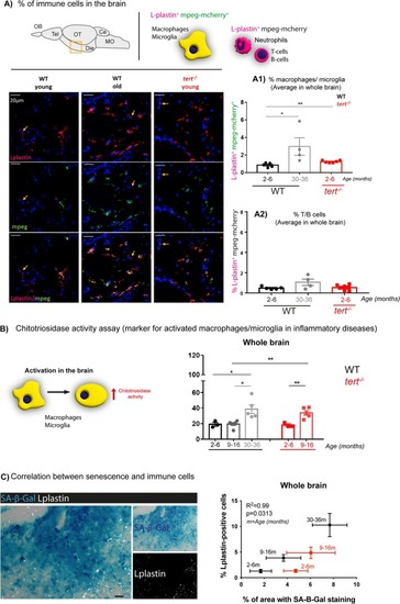
Telomerase depletion is associated with increased inflammation in the aged zebrafish brain. (A) Representative images of mpeg‐mcherry and L‐plastin staining in the diencephalon of adult zebrafish (schematic figure highlights the region imaged as diencephalon). The yellow arrows highlight Lplastin+; mpeg cells. (A1) Quantifications of L‐plastin‐positive; mpeg‐positive cells (orange; from red and green co‐localisation) and (A2) L‐plastin‐positive; mpeg‐negative cells (red) in the whole zebrafish brain show an increased number of macrophages/microglia (L‐plastin‐positive; mpeg‐positive cells) with natural ageing, and this is accelerated in the absence of telomerase (in tert−/− at 2–6 months of age). However, we observed no differences in the number of T/B cells and neutrophils (L‐plastin‐positive; mpeg‐negative cells) with ageing, in the WT or tert−/− fish. N = −6 per group. (B) Schematic figure of the chitotriosidase assay (left). Quantifications (right) show that chitotriosidase activity increases with natural ageing at > 30 months of age, and that this is accelerated in the tert−/− at the age of 9–16 months. (C) Representative images of co‐staining with SA‐β‐Gal (blue) and L‐plastin (white) in brain sections show that most of the L‐plastin‐positive cells do not co‐localise with the SA‐β‐Gal staining (left). Staining quantification shows a significant correlation between increased number of immune cells and increased expression of SA‐β‐Gal with ageing in both WT and tert−/− (right). (A1, A2, B) Each dot represents one animal. (A1, A2, B, C) Bar errors represent the SEM. * < 0.05; ** < 0.01.

Expression Data

Expression Detail
Antibody Labeling
Phenotype Data

Phenotype Detail
Acknowledgments
This image is the copyrighted work of the attributed author or publisher, and ZFIN has permission only to display this image to its users. Additional permissions should be obtained from the applicable author or publisher of the image. Full text @ Aging Cell