|
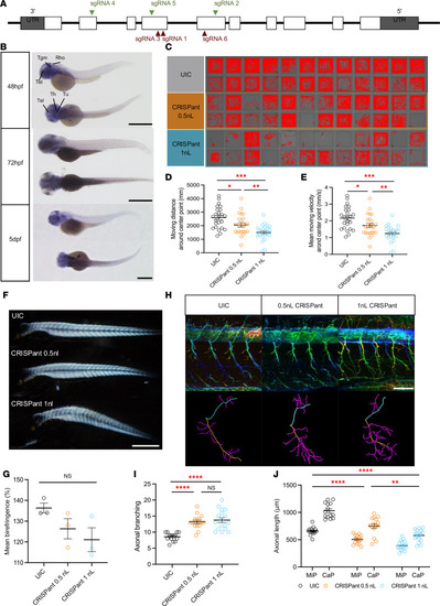
Zebrafish Danio rerioarhgap19 is important for motor neuron function. (A) Schematic representation of 10 exons to cover the complete coding region. The position of the sgRNA targets are indicated (green illustrate the highest level of knockdown efficiency). (B) Zebrafish arhgap19 expression at 3 different embryonic stages. At 48 hpf, WISH signal of arhgap19 is localized in the forebrain and hindbrain regions; scale bar: 1,000 μm. Rho, Rhombencephalon (hindbrain); Tel, Telencephalon; Th, Thalamus; Tu, Tuberculum; Tgm, Tegmentum. (C–E) Behavior analysis of UIC and arhgap19 mutant larvae at 5 dpf. C reveals the swimming trajectories of each larva. Quantification of total swimming distance (D) and swimming velocity (E) of UIC and arhgap19 mutant zebrafish larvae for 30 minutes (UIC, CRISPant 0.5 nL, CRISPant 1 nL: n = 24). Each bar represents mean (± SEM). Asterisks above the bars indicate significant difference (*P ≤ 0.05, **P ≤ 0.01, ***P ≤ 0.001). (F and G) UIC and arhgap19 mutant zebrafish larvae at 5 dpf were evaluated for muscle integrity using birefringence. (F) A representative image of one larva from each treatment group; scale bar: 1,000 μm. Each bar in plot G represents average birefringence (± SEM) for all zebrafish larvae (UIC, CRISPant 0.5 nL, CRISPant 1 nL: n = 3). (H–J) SMNs morphogenesis defects in arhgap19 mutant zebrafish larvae. (H) Confocal imaging analysis and 3D reconstruction of SMNs in UIC and arhgap19 mutant groups at 5 dpf; scale bar: 100 μm. (I) Axonal branching number of Cap axons in UIC and arhgap19 mutant zebrafish larvae. (J) Average axonal length of Cap (yellow) and Mip (blue) axons in UIC and arhgap19 mutant zebrafish larvae. Each bar represents mean (± SEM). Asterisks above the bars indicate significant difference (*P ≤ 0.05, **P ≤ 0.01, ***P ≤ 0.001, ****P ≤ 0.0001).
|