|
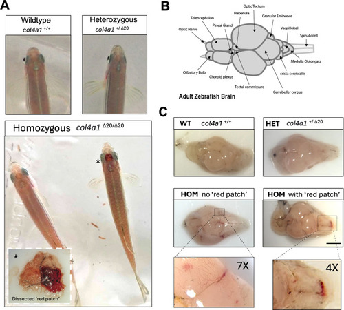
col4a1Δ20 mutant fish display spontaneous brain haemorrhages during adulthood. A. Two adult homozygous (col4a1Δ20/Δ20) zebrafish viewed dorsally with a cephalic ‘red patch’ (right, marked with an *) and one without (left) (lower panel). The dashed white outlined insert image is a representative image of a successfully dissected ‘red patch’. Above are the heads of wildtype (col4a1+/+) and heterozygous (col4a1+/Δ20) siblings. B. A diagram of the dorsal adult zebrafish brain adapted from [61] and created in Adobe Illustrator. C. Representative images of whole brains from adult sibling zebrafish at 12 months old. The top two panels are brains from col4a1+/+and col4a1+/Δ20fish and the bottom two panels are brains from col4a1Δ20/Δ20 fish with (right) and without (left) a visible ‘red patch’. The images below are magnifications of the regions indicated. The scale bar represents 1 mm.
|