
Fig. 3
- ID
- ZDB-FIG-251021-57
- Publication
- Gonzalez et al., 2025 - Synergistic and independent roles for Nodal and FGF in zebrafish CPC migration and asymmetric heart morphogenesis
- Other Figures
- All Figure Page
- Back to All Figure Page
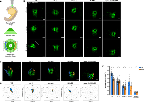
FGF signaling promotes CPC migration while Nodal promotes CPC migration and laterality during jogging. (A) Schematic of the imaging strategy for time-lapse imaging of the cardiac cone throughout jogging, with outer atrial cells in light green and inner ventricular cells in dark green. Left-sided (blue) and right-sided (orange) cells that underwent tracking for analysis are shown. (B) Representative time-lapse images depicting CPC migration from the formation of the cardiac cone through the next 3 h of development. WT and ntl−/− embryos typically complete jogging and tube formation in this time frame. Arrows indicate heart tube direction. Scale bars: 50 µm. (C) Trajectories of left-sided (blue) and right-sided (orange) CPCs during 3 h of jogging. Scale bars: 50 µm. (D) Rose plots demonstrating the angle of displacement of each tracked left-sided (blue) and right-sided (orange) CPC after 3 h of jogging. (E) Average velocity of left-sided and right-sided CPCs during 3 h of jogging. *P<0.05, ***P<0.0001 (unpaired two-tailed Student's t-test). ns, not significant. (C-E) n=3 embryos (five left-sided and five right-sided cells tracked per embryo) per condition. Error bars represent s.d. (B,C) Images are dorsal views, with anterior to the top and left to the reader's left. |