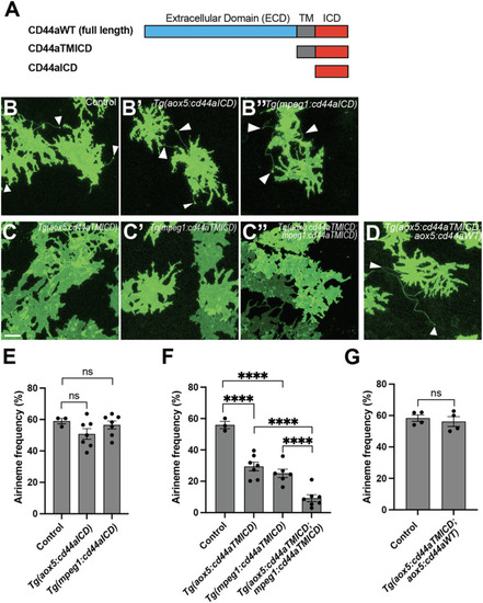
The crucial role of the extracellular domain of CD44 in airineme extension. (A) The schematics indicate the structure of CD44aWT, CD44aTMICD, and CD44aICD. (B–B″) Representative images of xanthophores extending airinemes in control, Tg(aox5:cd44aICD), or Tg(mpeg1:cd44aICD). (C–C″) Representative images of xanthophores in Tg(aox5:cd44aTMICD), Tg(mpeg1:cd44aTMICD), or Tg(aox5:cd44aTMICD; mpeg1:cd44aTMICD). (D) Representative image of xanthophores in Tg(aox5:cd44aTMICD; aox5:cd44aWT). White arrowheads mark airinemes (E) Airineme extension frequency was not significantly altered in transgenic embryos that overexpressed the intracellular domain of cd44 (cd44aICD) specifically in the xanthophore-lineages or macrophages, (F (2, 14) = 1.736, P = 0.2121, 17 embryos in total). (F) Overexpression of CD44a with a truncated extracellular domain (cd44aTMICD) exhibited a significant reduction in airineme extension frequency in both the xanthophore-lineages and macrophages, (F (2, 13) = 23.62, P < 0.0001, 16 embryos in total). Airineme extension frequency was further significantly reduced when cd44aTMICD was overexpressed in both cell type simultaneously, (F (2, 17) = 18.04, P < 0.0001). (G) Airineme extension frequency was restored when WT CD44 was overexpressed in embryos already expressing cd44aTMICD in the xanthophore-lineages, (P = 0.5767, 4 embryos each). Statistically significant results were evaluated using a one-way ANOVA, followed by Tukey’s HSD post hoc test or Student’s t-test. Error bars indicate mean ± SEM.
|
