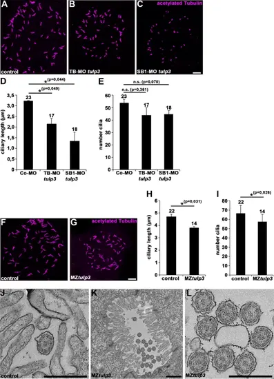
Tulp3 deficiency results in defective cilia formation and function in zebrafish. (A-C) Representative confocal images of the Kupffer’s vesicle of Co-MO (2ng) (A), TB-MO tulp3 (2ng) (B) or SB1-MO tulp3 (0,5ng) (C) injected embryos at 8 somites immunostained with anti-acetylated Tubulin as a ciliary marker. Scale bar: 10 μm. (D, E) Quantification of the ciliary length (D) and cilia number (E) in the Kupffer’s vesicle of Co-MO (3 independent experiments; n = 9, n = 7 and n = 7 analysed embryos, respectively), TB-MO tulp3 (n = 6, n = 4 and n = 7) or SB1-MO tulp3 (n = 4, n = 6 and n = 8) injected embryos at 8 somites; total number of embryos used for analyses are shown above respective bar. (F, G) Representative confocal images of the Kupffer’s vesicle of MZtulp3 embryo (G) and respective control embryo (F) at 8 somites immunostained with anti-acetylated Tubulin. Scale bar: 10 μm. (H, I) Quantification of the ciliary length (H) and cilia number (I) in the Kupffer’s vesicle of control embryos (3 independent experiments; n = 7, n = 7 and n = 8 analysed embryos, respectively) and MZtulp3 embryos (n = 6, n = 4 and n = 4) at 8 somites; total number of embryos used for analyses are shown above respective bar. (J-L) TEM of pronephric tubule microvilli and motile cilia of control embryos (J) and MZtulp3 (K, L) embryos (with severe ventral body curvature) at 5dpf. Scale bars: 500 nm (J, L); 1 μm (K).
|
